
|
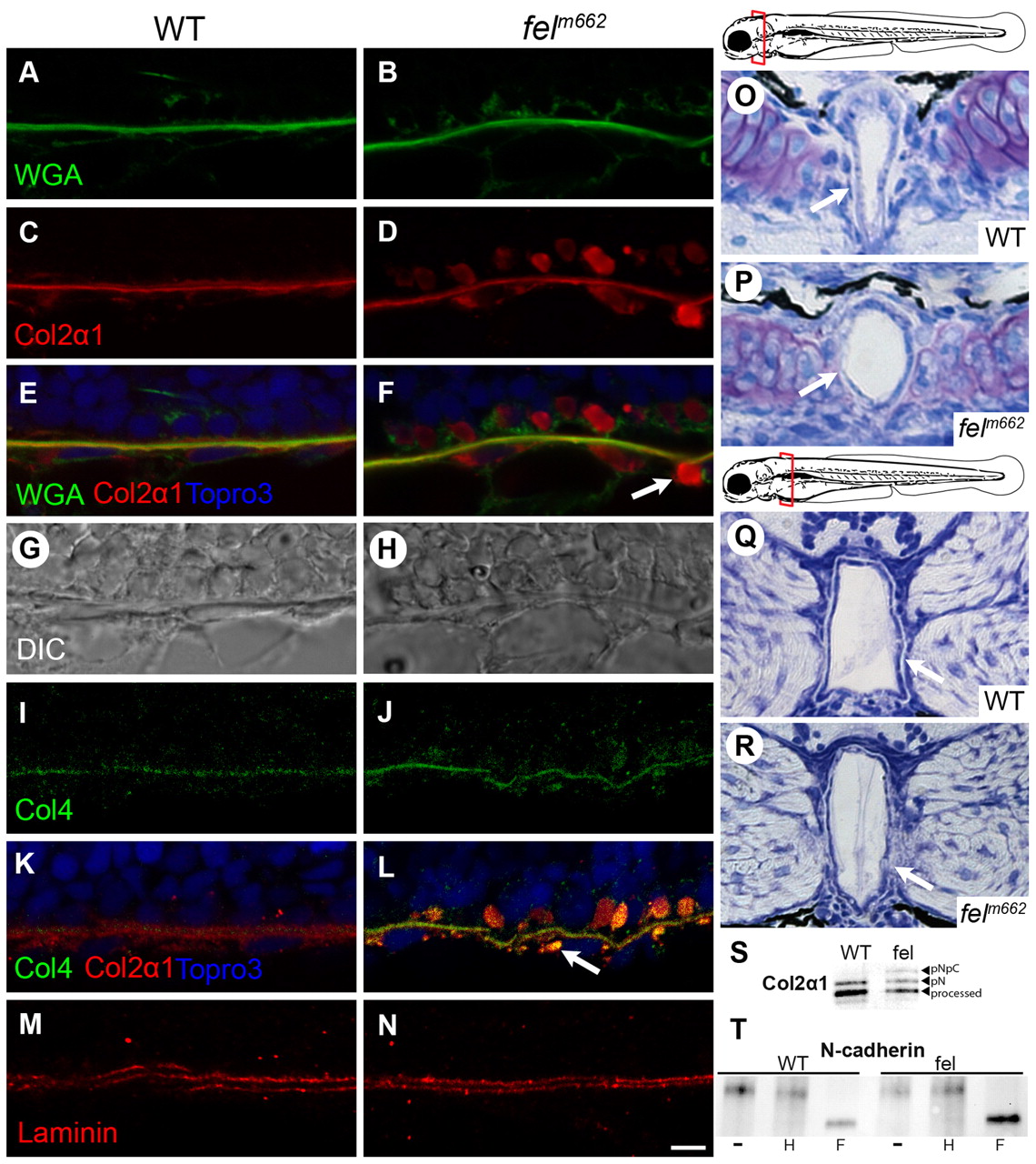
Fig. 3
Collagen trafficking is preferentially disrupted in feelgood mutants, leading to notochord defects. (A–F) Immunofluorescence of WGA, Col2α1 and merged images of 15-µm sagittal sections of the notochord of 28-hpf embryos. Arrow indicates vesicle-like collagen staining outside the notochord sheath. (G,H) DIC images of the corresponding sections in A–F. (I–N) Immunofluorescence of type-IV and type-II collagen (I–L) and laminin (M,N) in 15-μm sagittal sections of the notochord at 28 hpf. Scale bar: 5 μm. (O–R) Toluidine blue staining of transverse sections through the notochord at the level of the posterior parachordal plate (O,P) and posterior medulla oblongata (Q,R) in 80-hpf embryos. Schematic diagrams mark the plane of the corresponding sections. Arrows indicate the less robust notochord sheath in feelgood compared with wild types. (S) Immunoblot analysis of type II collagen processing. Molecular forms are indicated as: processed, the fully processed form; pN, pN-collagen II; pNpC, unprocessed procollagen II. (T) EndoH sensitivity assay for N-cadherin. Embryo lysates were either untreated (–) or treated with either EndoH (H) or PNGase F (F) before immunoblotting for N-cadherin.