Fig. 3
Fig. 3
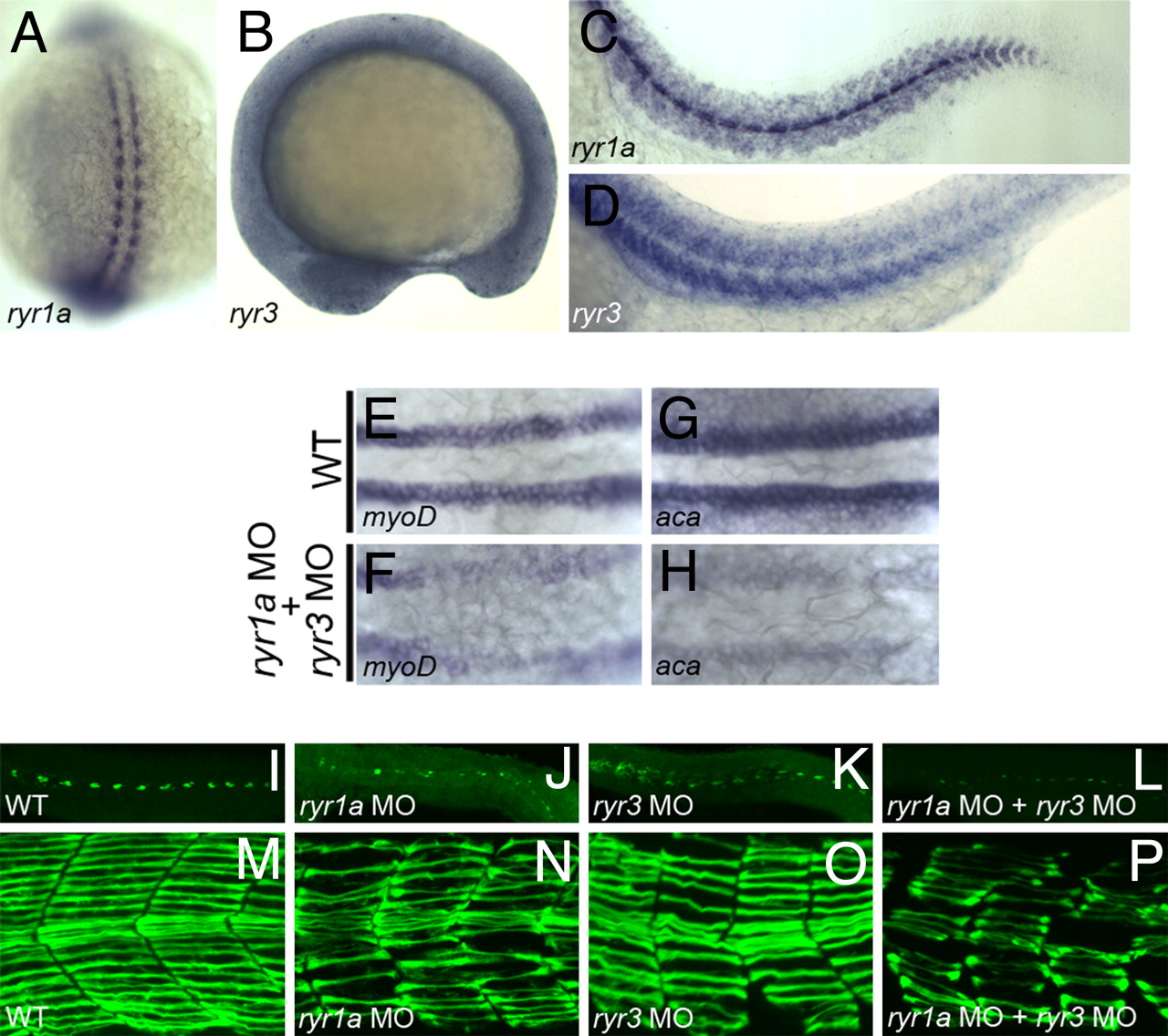
Ryanodine receptors and SepN have similar functions in muscle formation. (A–D) Expression of ryr1a and ryr3 transcripts in 15-somite (A and B) and 24 hpf (C and D) embryos. (E–H) Expression of the myogenic lineage genes, myoD and α-cardiac actin, is greatly reduced in the adaxial cell population of ryr1a; ryr3 double morphants. (I–L) 4D9+ slow muscle pioneer fiber nuclei and (M–P) F59+ slow muscle fibers are reduced in number and irregularly shaped in 24-hpf embryos depleted for RyR1a, RyR3, or both RyRs. (A) Dorsal view, rostral up. B–D and I–P, lateral views, rostral left. (E–H) Dorsal views, rostral left.

