
|
Fig. 3
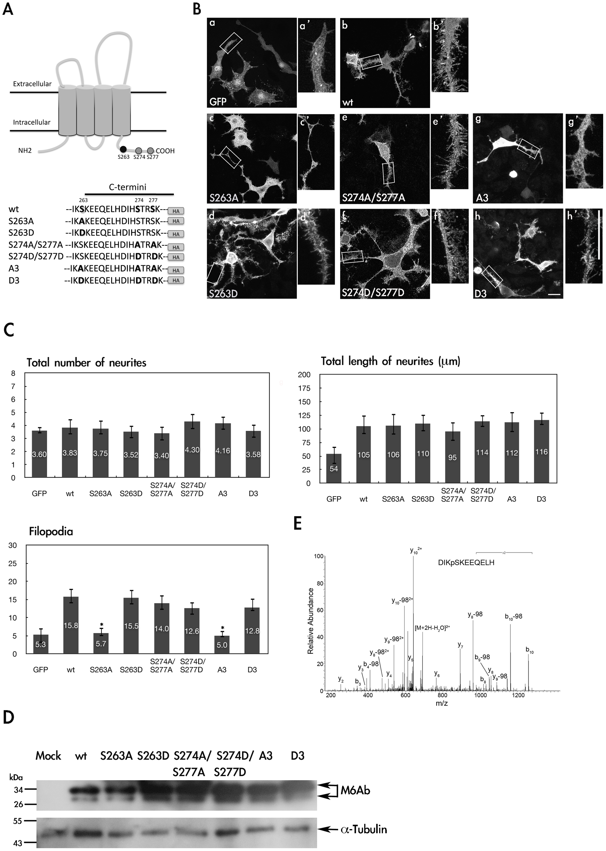
Phosphorylation of S263 is critical for regulation of filopodium formation in PC12 cells.
(A) Partial amino acid sequences of the wide-type and mutant proteins of zebrafish M6Ab. (B) PC12 cells were transfected with pcDNA3-GFP-HA(a), pcDNA3-M6Ab-HA(b), pcDNA3-M6Ab(S263A)-HA(c), pcDNA3-M6Ab (S263D)-HA(d), pcDNA3-M6Ab(S274A/S277A)-HA(e), pcDNA3-M6Ab(S274D/S277D)-HA(f), pcDNA3-M6Ab(A3)-HA(g), or pcDNA3-M6Ab(D3)-HA(h) plasmids. Twenty-four hours after transfection, cells were treated with nerve growth factor (NGF) (100 ng/ml) for 2 days. Then transfected cells were fixed, and M6Ab-HA was detected using anti-HA antibodies for immunostaining. PC12 neurites are shown at a higher magnification (a2–h2). (C) Quantification of the total number of neurites, total length of neurites, and filopodium-like processes in a 20- · m neurite length. Results are expressed as the mean ± SD of at least 40~50 neurites. At least three independent experiments were analyzed. *Significant difference compared with the respective control of WT-M6Ab overexpression (P<0.05). (D) Cell lysates from different transfected cells as indicated were extracted and immunoblotted with an anti-HA antibody or anti-Tubulin antibody. (E) MS/MS spectrum on [M+2H]2+ (m/z 718.32) ion for the peptides DIKpSKEEQELH from WT M6Ab protein. The product ion y8 which carries a phosphate indicated that Serine 263 was phosphorylated. Residues bearing phosphate moieties are indicated with p. “b” and “y” ion series represent fragment ions containing the N- and C-termini of the peptide, respectively. Scale bars, 10 μm.