
|
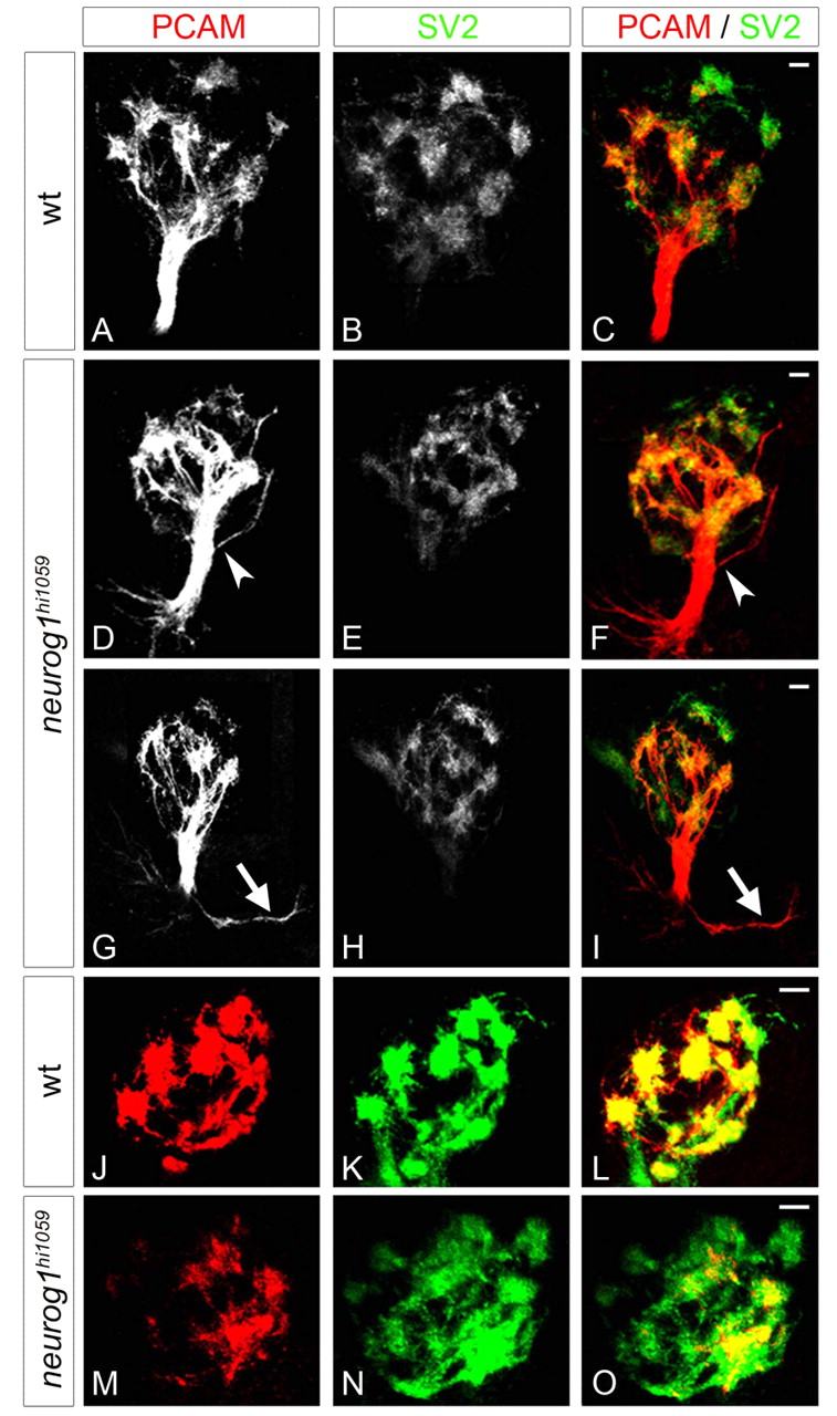
Fig. 8
The fasciculation of olfactory axons and organisation of proto-glomeruli are flawed in neurog1 mutant zebrafish embryos. (A-I) Confocal projections of immunolabelling against PCAM and SV2 of olfactory projections in wild-type (A-C) and neurog1 mutant (D-I) embryos at 72 hpf. Whereas projections in wild-type embryos form a single fasciculated tract from the olfactory placode to the olfactory bulb, fasciculation (arrowheads in D,F) is aberrant in neurog1 mutant embryos; certain mutant embryos display abnormal guidance of projections immediately after they leave the placode (arrows in G,I). (J-O) Confocal projections of immunolabelling against PCAM and SV2 in the olfactory bulb in wild-type (J-L) and neurog1 mutant (M-O) embryos at 72 hpf. Proto-glomeruli appear less well organised in neurog1 mutant embryos relative to wild-type siblings. Embryos are viewed frontally (A-I) or dorsally with anterior up (J-O). Scale bars: 10 μM.