
|
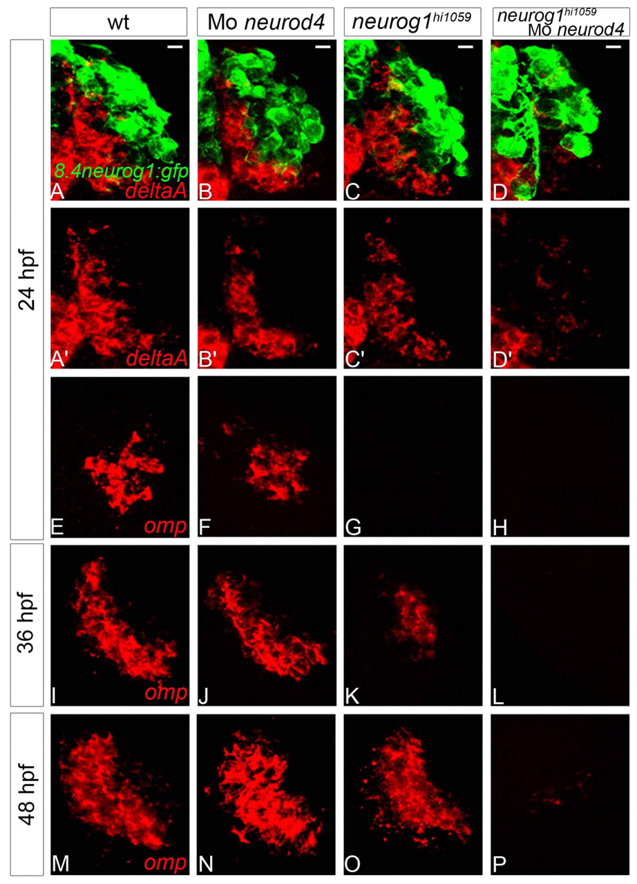
Fig. 6
Neurod4 and Neurog1 act redundantly during OSN development in zebrafish. (A-D2) Confocal projections of in situ hybridisation and immunolabelling against deltaA and GFP in wild-type (A,A2), neurod4 morphant (B,B2), neurog1 mutant (C,C2) and neurog1/neurod4 double loss-of-function (D,D2) olfactory placodes of Tg(8.4neurog1:GFP) embryos at 24 hpf. Although no difference is apparent in the expression of deltaA in single loss-of-function embryos relative to wild-type siblings, deltaA expression is absent when both neurog1 and neurod4 function is abolished. (E-P) Confocal sections of in situ hybridisation labelling against olfactory marker protein (omp) in wild-type (E,I,M), neurod4 morphant (F,J,N), neurog1 mutant (G,K,O) and neurog1/neurod4 double loss-of-function (H,L,P) olfactory placodes at 24 (E-H), 36 (I-L) and 48 (M-P) hpf. Although omp expression resembles the wild-type pattern in the single loss-of-function context at 48 hpf, there is a delay in the initiation of omp expression in the absence of Neurog1 function. The expression of omp is never detected when both neurog1 and neurod4 function is abolished. Scale bars: 10 μm.