
Image
|
Figure Caption
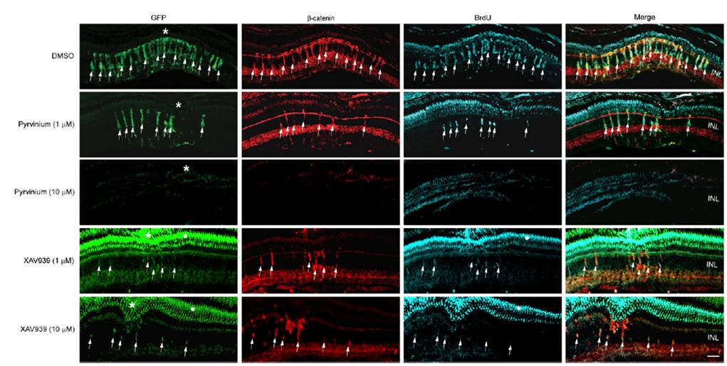
Fig. S5 β-catenin destabilization inhibits MG dedifferentiation and proliferation in the injured retina. Retinas in 1016 tuba1a:gfp transgenic fish were injured and injected with either DMSO, pyrvinium, or XAV939. Pyrvinium and XAV939 inhibited GFP expression, β-catenin stabilization, and cell proliferation. Asterisks indicate injury sites. White dot identifies areas of GFP autofluorescence that also bleeds over into the blue channel, due to the long exposures that are necessary to visualize cells harboring very low levels of GFP. INL, inner nuclear layer. (Scale bar, 20 μm.)
Acknowledgments
This image is the copyrighted work of the attributed author or publisher, and
ZFIN has permission only to display this image to its users.
Additional permissions should be obtained from the applicable author or publisher of the image.
Full text @ Proc. Natl. Acad. Sci. USA