
|
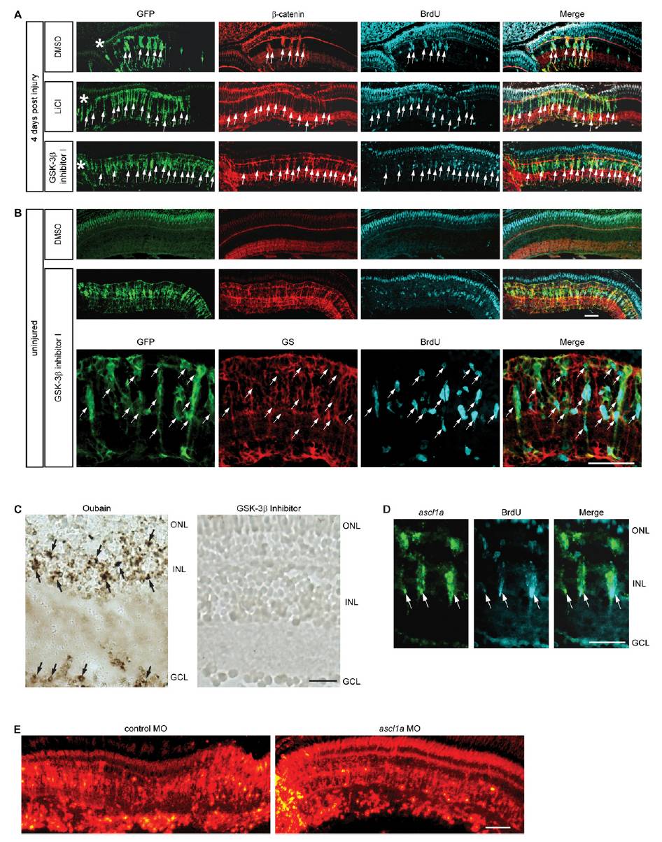
Fig. S6 Pharmacological stabilization of β-catenin stimulates MG dedifferentiation and proliferation in the injured and uninjured retina. (A) LiCl and GSK-3β inhibitor injection into the eye at the time of retinal injury increases the regenerative zone as indicated by the increased number of GFP transgene expressing cells and the increased number of cells labeled with BrdU in 1016 tuba1a:gfp transgenic fish. (B) Intravitreal injection of the GSK-3β inhibitor into uninjured eyes stimulates MG dedifferentiation and cell proliferation in 1016 tuba1a:gfp transgenic fish examined at 4 d postinjection. Upper and Middle are a low magnification view of the retina; Bottom is a close-up view to show that glutamine synthetase (GS)-expressing MG have dedifferentiated (GFP+) and begun to proliferate (BrdU+) in response to GSK-3β inhibition. Asterisks indicate the injury sites. (C) TUNEL stain shows a lack of cell death in GSK-3β inhibitor-treated retinas. As a positive control retinas were treated with oubain, which resulted in widespread cell death. (D) In situ hybridization and BrdU immunofluorescence show GSK-3β inhibition-dependent induction of ascl1a in the uninjured retina is in proliferating MG-derived progenitors. (E) Retinal sections showing lissamine- labeled control and ascl1a-targeting MOs are widely incorporated into most cells of the retina. ONL, outer nuclear layer; INL, inner nuclear layer; GCL, ganglion cell layer. (Scale bars, 20 μm.)