
|
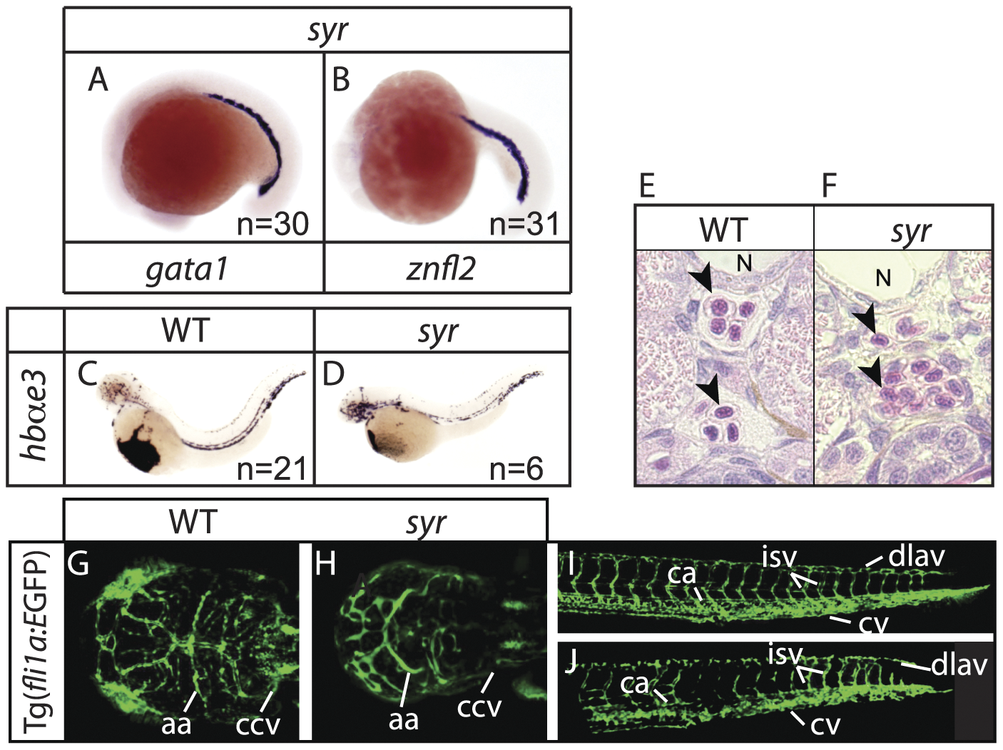
Fig. 4
Erythropoiesis proceeds normally in syr.
Examination of early erythroid markers at 17–19 hpf in syr (A–B); staining of embryonic globin in wt and syr at 48 hpf (C–D); transverse sections of WT and syr embryos at 3 dpf, counterstained with hematoxylin and eosin; notochord (N) and neutrophils (arrowheads) are indicated (E–F); syr crossed with Tg(fli1a:EGFP) shows close to normal vasculature in both head (G–H) and tail (I–J). Heads are dorsal view with anterior to left, WT on the left (G) and syr on the right (H); aa (aortic arches) and ccv (common cardinal vein) are indicated. Tails are lateral view, anterior to left and ca (caudal artery), cv (caudal vein), dlav (dorsal longitudinal anastomotic vessel) and isv (intersomitic vessels) are indicated.