
Fig. 5
|
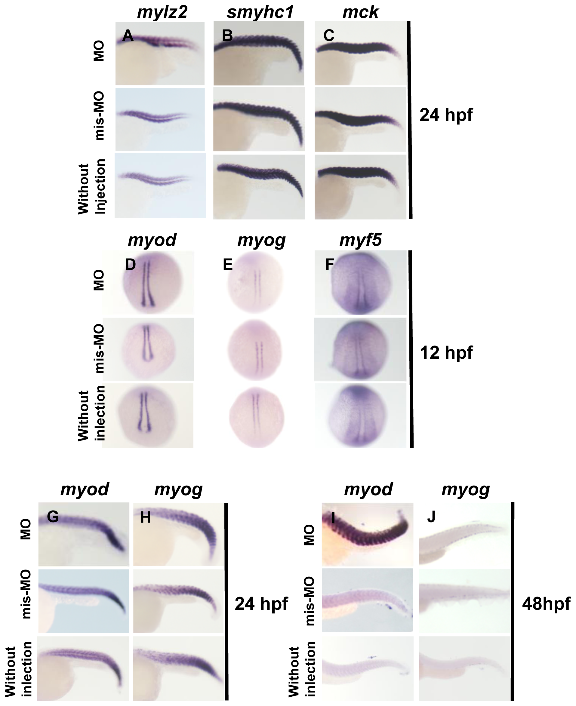
Fig. 5
In situ hybridization analysis of terminal differentiation markers of skeletal muscle and myogenic regulatory factors.
(A, B, and C) Expression of mylz2, smyhc1 and mck in Smyd3 morphants, control embryos injected with Smyd3-mis-MO and without injection at 24 hpf. (D, E, F, G, H, I, and J) Expression of myod, myog, and myf5 in Smyd3 morphants, control embryos injected with Smyd3-mis-MO and without injection at 12 hpf (D, E, and F), 24 hpf (G and H) and 48 hpf (I and J). Embryos are shown in lateral view, anterior toward the left (A, B, C, G, H, I, and J). Embryos are shown in dorsal view, anterior toward the top (D, E, and F).