
|
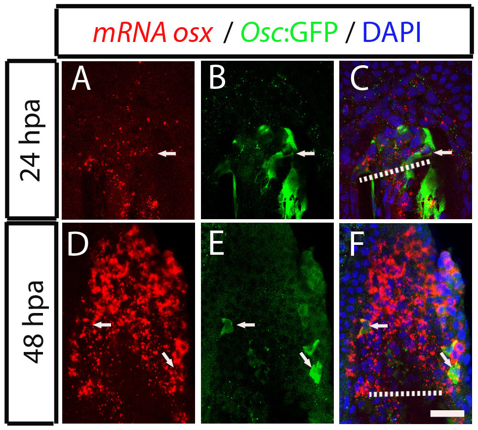
Fig. S5
Dedifferentiating osteocalcin-positive scleroblasts at the blastema start to express de novo osterix at 48 hpa. (A-F) Osterix mRNA expression detected by fluorescence in situ hybridization followed by immunohistochemistry against GFP in Tg(osteocalcin:GFP) fins. (A-C) 24 hpa fin. The arrow indicates a osteocalcin-positive cell at the blastema that does not express osterix mRNA, labelled with Fast Red. (D-F) 48 hpa fin. The arrows indicate double positive cells co-expressing osterix mRNA and osteocalcin:GFP. The images are each projections of several confocal optical slices. Scale bar: 50 µm. Dashed lines indicate amputation plane.