
Image
|
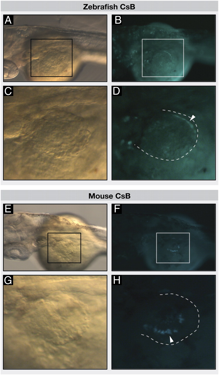
Figure Caption
Fig. 2
Vertebrate CsB enhancers drive neural tube and distal fin GFP expression in transgenic zebrafish. Transgenic zebrafish embryos injected with zebrafish CsB (A–D) or mouse CsB (E–H), at 52 hpf, anterior to the left. Dorsal view of zebrafish embryos; black and white boxes indicate position of the fin in bright field (A and E) and fluorescence (B and F), respectively. (C and G) Bright-field images show dorsal view of fins. (D and H) Dotted lines indicate position of fins, white arrowheads indicate eGFP signal in fins.
Acknowledgments
This image is the copyrighted work of the attributed author or publisher, and
ZFIN has permission only to display this image to its users.
Additional permissions should be obtained from the applicable author or publisher of the image.
Full text @ Proc. Natl. Acad. Sci. USA