
Fig. S1
- ID
- ZDB-IMAGE-110812-41
- Genes
- Publication
- Lim et al., 2011 - Motoneurons are essential for vascular pathfinding
- All Figures
- Figures for Lim et al., 2011
|
Fig. S1
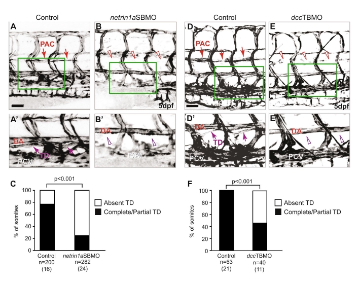
netrin 1a and dcc morphants do not form a complete thoracic duct. (A,B,D,E) 5 dpf fli1a:egfpy1 larvae; reversed contrast, confocal projections. (A2,B2,D2,E2) Enlargements of the boxed regions from A,B,D,E. (A-A2,D-D2) The thoracic duct (TD) forms normally (arrows) between the dorsal aorta (DA) and posterior cardinal vein (PCV) in uninjected control embryos. (B,B2,E,E2) The TD is absent in netrin1aSBMO or dccTBMO morphants (open arrowheads). (C,F) Quantitation of partial or complete TD formation in control embryos (77%), netrin1aSBMO morphants (25%) and dccTBMO morphants (45±19%; mean±s.e.m.); P-values, Mann-Whitney U test. n, number of hemisegments (number of embryos). PAC, parachordal chain; DA, dorsal aorta; PCV, posterior cardinal vein. Scale bars: 50 μm.