
|
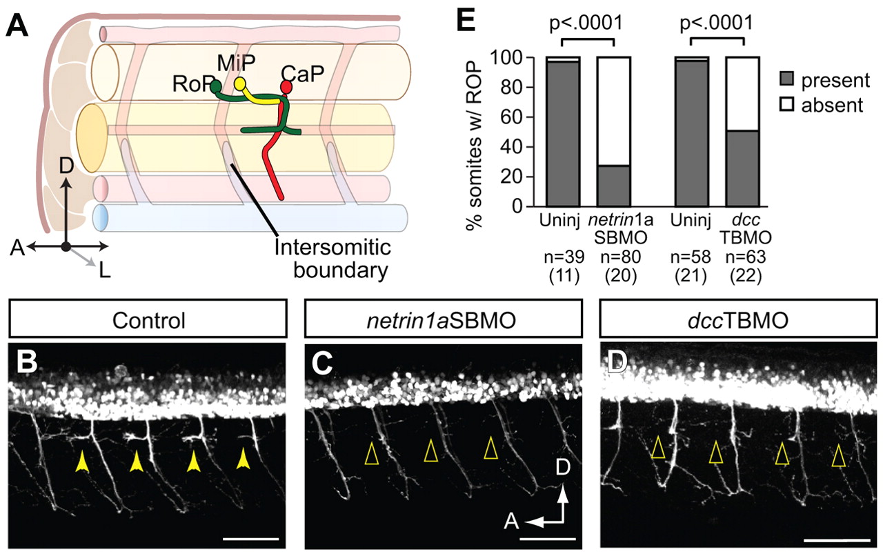
Fig. 6
RoP axons and associated secondary axons at the HMS do not form in netrin 1a and dcc morphants. (A) Diagram of RoP, middle primary (MiP) and caudal primary (CaP) axons at 36 hpf. (B-D) Lateral view of 55 hpf hb9:GFP zebrafish embryos injected with (B) no MO, (C) netrin 1a splice-blocking MO (netrin1aSBMO) or (D) dcc translation-blocking MO (dccTBMO). Confocal projections. (B) Uninjected controls show axons at the HMS in almost every somite (arrowheads). In embryos injected with (C) netrin1aSBMO or (D) dccTBMO, axons fail to form at the HMS (arrowheads). (E) Axons at the HMS were counted in 3-4 somites per embryo in segments 7-11 in control, netrin1aSBMO- and dccTBMO-injected embryos. Uninjected, 97±3%; netrin 1a morphant, 27±7%. Uninjected, 98±2%; dcc morphant, 49±7%. All values are mean±s.e.m.; P-value determined by Mann-Whitney U test. n, number of hemisegments (number of embryos). A, anterior; D, dorsal; L, lateral; RoP, rostral primary motoneuron; MiP, middle primary motoneuron; CaP, caudal primary motoneuron. Scale bars: 50 μm.