
|
Fig. 1
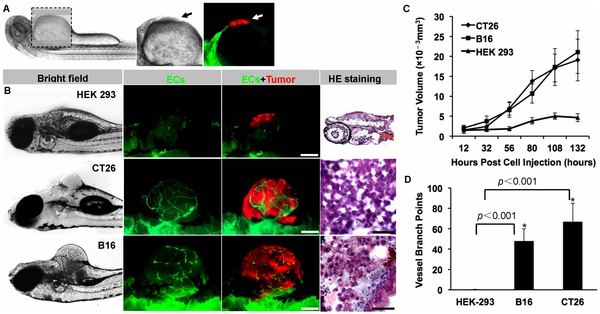
The establishment of mammalian tumor xenograft models in zebrafish.
Red fluorescence-labeled malignant tumor cells (murine colon carcinoma CT26 and melanoma B16 cells) and non-tumorigenic HEK 293 cells were implanted into the abdominal perivitelline space (A, indicated by arrow) of 48 hpf Tg(flk1:EGFP) transgenic zebrafish embryos (50–100 cells/embryo). When tumors (B, red) reached 350–450 μm in diameter when filled with neo-vessels (B, green), the neoplasia were isolated and analyzed by H&E staining (B). The growth rate of xenografted tumors (C) and the number of tumor-induced neovessels (D) were analyzed quantitatively. * indicates the significant difference. Scale bar, 100 μm.