
Fig. 3
|
Fig. 3
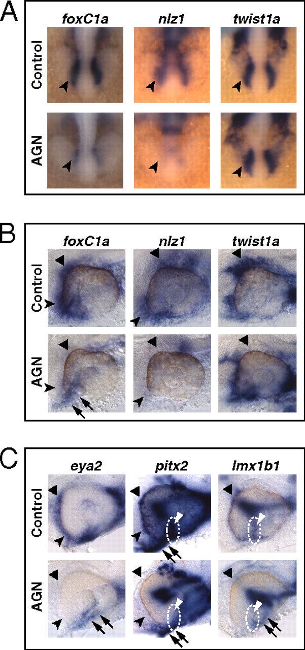
RAR signaling regulates gene expression in POM. (A–C) Embryos treated with DMSO or 10 μM AGN from 3s and hybridized at 18s (A), 24–25 hpf (B), or 31 hpf (C) with the indicated probes. (A) Dorsal/anterior views. In the AGN-treated embryos, foxC1a and nlz1, but not twist1a, are down-regulated in migrating POM (arrowheads). (B and C) Lateral views of eyes. AGN treatment decreases foxC1a, nlz1, eya2, pitx2, and lmx1b1 expression in both anterior-ventral (arrowheads) and anterior-dorsal (black triangles) POM. AGN treatment decreases twist1a expression in dorsal POM (black triangles). Expression of pitx2 and lmx1b1 within the choroid fissure is also diminished (white dashed circles and triangles). Residual foxC1a, eya2, and lmx1b1 staining is detectable in a ventro-medial location behind the eye (arrows). pitx2 is expressed in extraocular mesodermal tissue in both the control and the AGN-treated embryo (arrows). Panels are representative images of the following numbers of independent experiments: pitx2, n = 4; foxC1a, n = 3; and nlz1, twist1a, eya2, lmx1b1, n = 2. Approximately 10–30 embryos were assayed for each condition and probe in each experiment.