
|
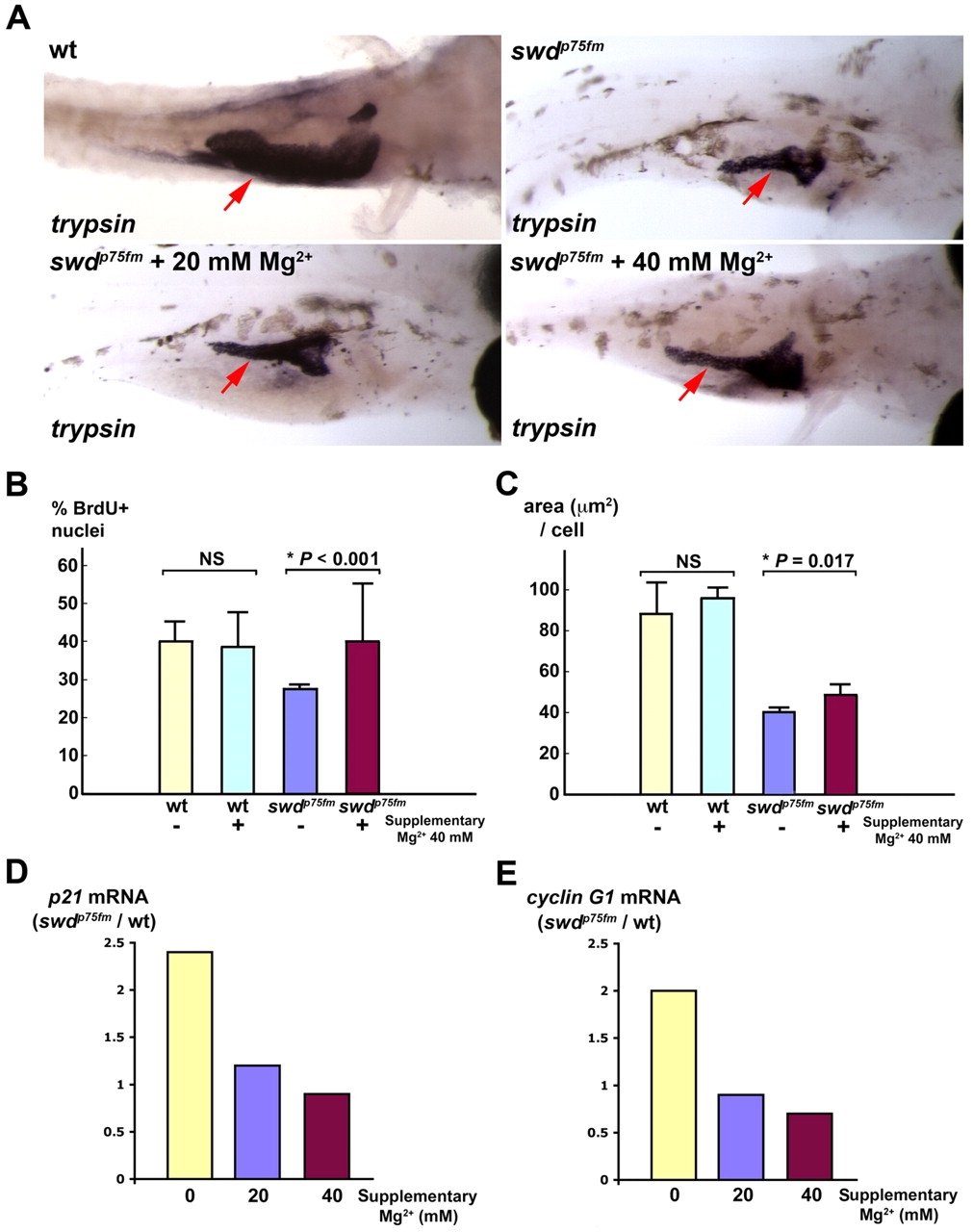
Fig. 5
Supplementary Mg2+ partially rescues the growth defect of exocrine pancreas in swd mutants by improving cell-cycle progression and cell growth, with repression of p21cdkn1a and cyclin G1. Exocrine pancreas of the swdp75fm mutants and WT siblings incubated in medium with or without supplementary 20 mM or 40 mM MgCl2 was analyzed. The swdp75fm mutants were identified on the basis of their hypopigmented skin, which was minimally affected by the supplementary MgCl2. (A) Exocrine pancreas (red arrows) in the larvae of 5 dpf embryos by in situ hybridization using anti-trypsin riboprobes. The WT embryos were grown in medium supplemented with PTU, which inhibits skin pigmentation and facilitates visualization of the trypsin-expressing exocrine pancreas. Each larva shown is representative of 40 larvae in each experimental group, and this experiment was performed three times with similar results. (B) Proliferation assay to determine the proportion of BrdU+ nuclei (cells in S phase) in the exocrine pancreas at 72 hpf. (C) Morphometric analysis of exocrine pancreatic cell growth (area, in μm2, per cell) at 5 dpf. Each value represents the mean of five larvae + s.d. *P<0.05 is considered statistically significant. NS, not statistically significant. (D,E) Relative mRNA levels of p21cdkn1a and cyclin G1 by quantitative real-time PCR at 5 dpf. Each value represents the ratio of p21cdkn1a or cyclin G1 mRNA in the swd mutants to WT in the same experimental group. This experiment was repeated with reproducible results.