
|
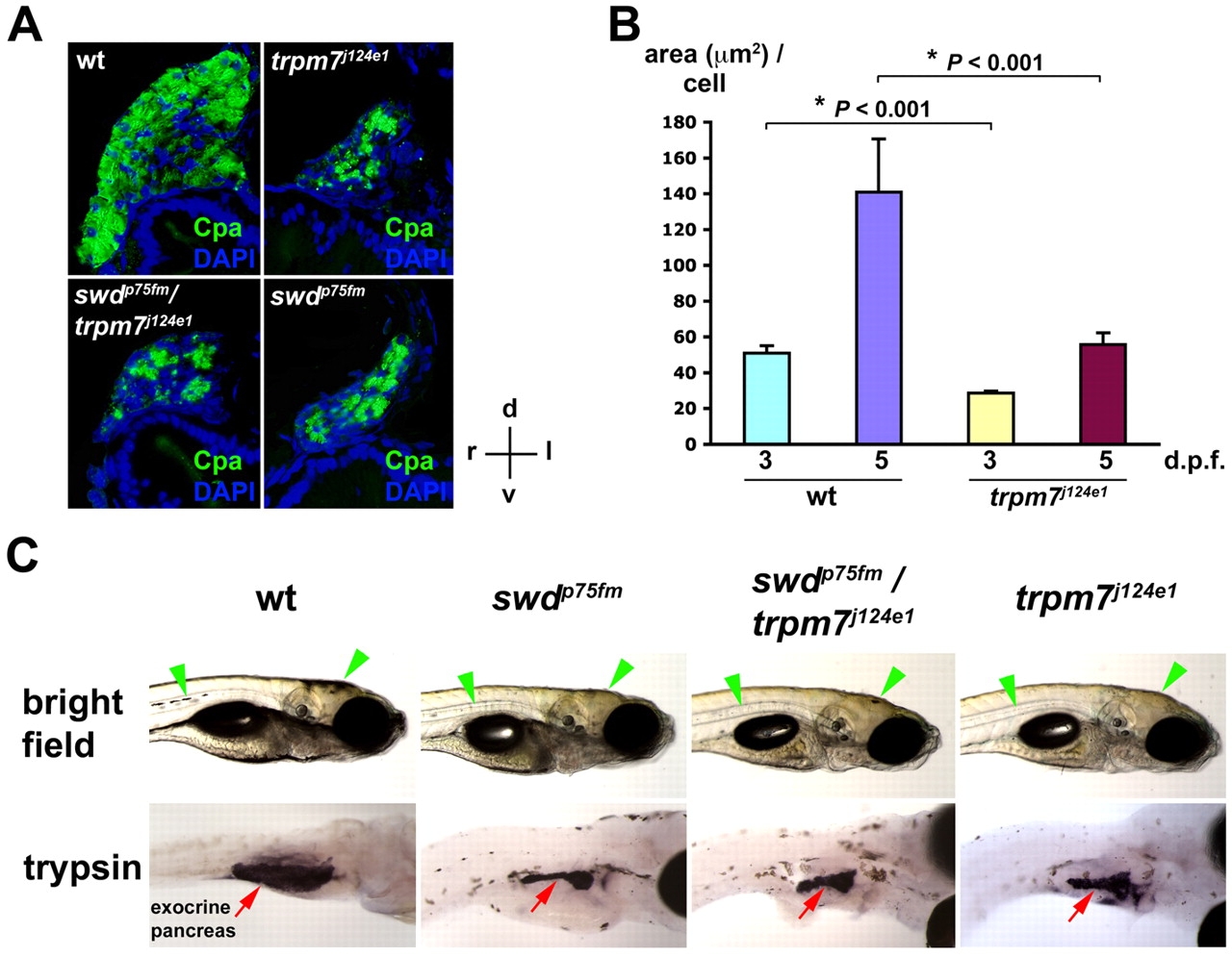
Fig. 3
The swd locus is allelic with the trpm7 gene. (A) Exocrine pancreas of WT, the trpm7j124e1 mutant, the swdp75fm/trpm7j124e1 mutant and the swdp75fm mutant on 5 dpf by immunohistochemistry using anti-Cpa antibodies followed by cross-sectional histological analysis. Staining with DAPI was used to visualize the nuclei. The histological sections are oriented as indicated: d, dorsal; v, ventral; r, right; l, left. (B) Morphometric analysis of exocrine pancreatic epithelial growth (area, in μm2, per cell) in the trpm7j124e1 mutants and WT siblings on 3 and 5 dpf. Each value represents the mean + s.e.m. Statistical analysis was performed using Student’s t-test, with *P<0.05 considered statistically significant. (C) Bright-field images in right lateral view and whole-mount in situ hybridization using anti-trypsin riboprobes. The swdp75fm/+ and trpm7j124e1/+ heterozygotes were crossed with each other, and the progeny larvae were analyzed on 5 dpf. The WT, but not mutant, larvae were grown in medium supplemented with PTU, which inhibits skin pigmentation, in order to facilitate visualization of the exocrine-pancreas-expressing trypsin. Note the regions of skin pigmentation (green arrowheads) and trypsin-expressing exocrine pancreas (red arrows) in the mutants as compared to WT.