
|
Fig. 6
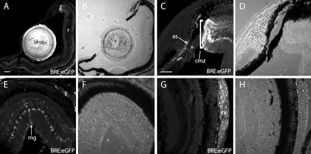
Figure 7. Adult ocular bone morphogenic protein response element:enhanced green fluorescent protein (BRE:eGFP) expression revealed in cryosections. A: BRE:eGFP expression was obvious in the lens and dorsal ciliary margin, but not in the central retina (r). B: Transmitted light image of A. C: BRE:eGFP expression in the ciliary marginal zone (cmz) and anterior stromal and vascular cells (as, arrows). D: Transmitted light image of C. E: BRE:eGFP expression in dorsal retina showing Müller glia (mg) and scleral cells (s). F: Transmitted light image of E. G: BRE:eGFP expression in central retina showing choroidal rete vascular cells (cv). Note the lack of glial expression in the central retina. H: Transmitted light image of G. Scale bars = 200 μm in A,B, 50 μm in C–H