
|
Fig. 3
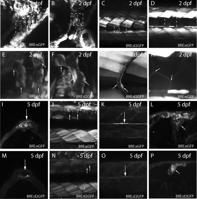
Figure 4. Diverse tissue expression of enhanced green fluorescent protein (eGFP) or destabilized eGFP (d2GFP) in bone morphogenic protein response element (BRE) transgenic lines. A: Expression of eGFP in cranial vasculature of 2 day postfertilization (dpf) BRE:eGFP larvae. B: Similar expression of d2GFP in 2 dpf cranial vasculature of BRE:d2GFP larvae. C: Expression of d2GFP in intersomitic vasculature of 2 dpf BRE:d2GFP larvae. D: Expression of eGFP in 2 dpf somite muscle cells and underlying notochord cells of BRE:eGFP larvae. E–H: Higher magnification of boxed regions in A–D. Arrows indicate the vascular structures in E, F, and G, and the somite muscle cells in H. I: Expression of eGFP in pineal cells of 5 dpf BRE:eGFP larvae (arrow). J: Expression of eGFP in somite muscle (s) and dorsal spinal neurons (arrows) of 5 dpf BRE:eGFP larvae. K: Expression of eGFP in medial longitudinal fasciculus axons (arrow) of 5 dpf BRE:eGFP larvae. L: Expression of eGFP in midbrain–hindbrain neurons (arrow) of 5 dpf BRE:eGFP larvae. Note the ependymal cells that are also weakly eGFP-positive (asterisks). M: Expression of d2GFP in pineal cells of 5 dpf BRE:d2GFP larvae (arrow). N: Expression of d2GFP in somite muscle (s) and dorsal spinal neurons (arrows) of 5 dpf BRE:eGFP larvae. O: Expression of d2GFP in medial longitudinal fasciculus axons (arrow) of 5 dpf BRE:d2GFP larvae. P: Expression of d2GFP in hindbrain neural progenitor cells (arrow) of 5 dpf BRE:d2GFP larvae.