
|
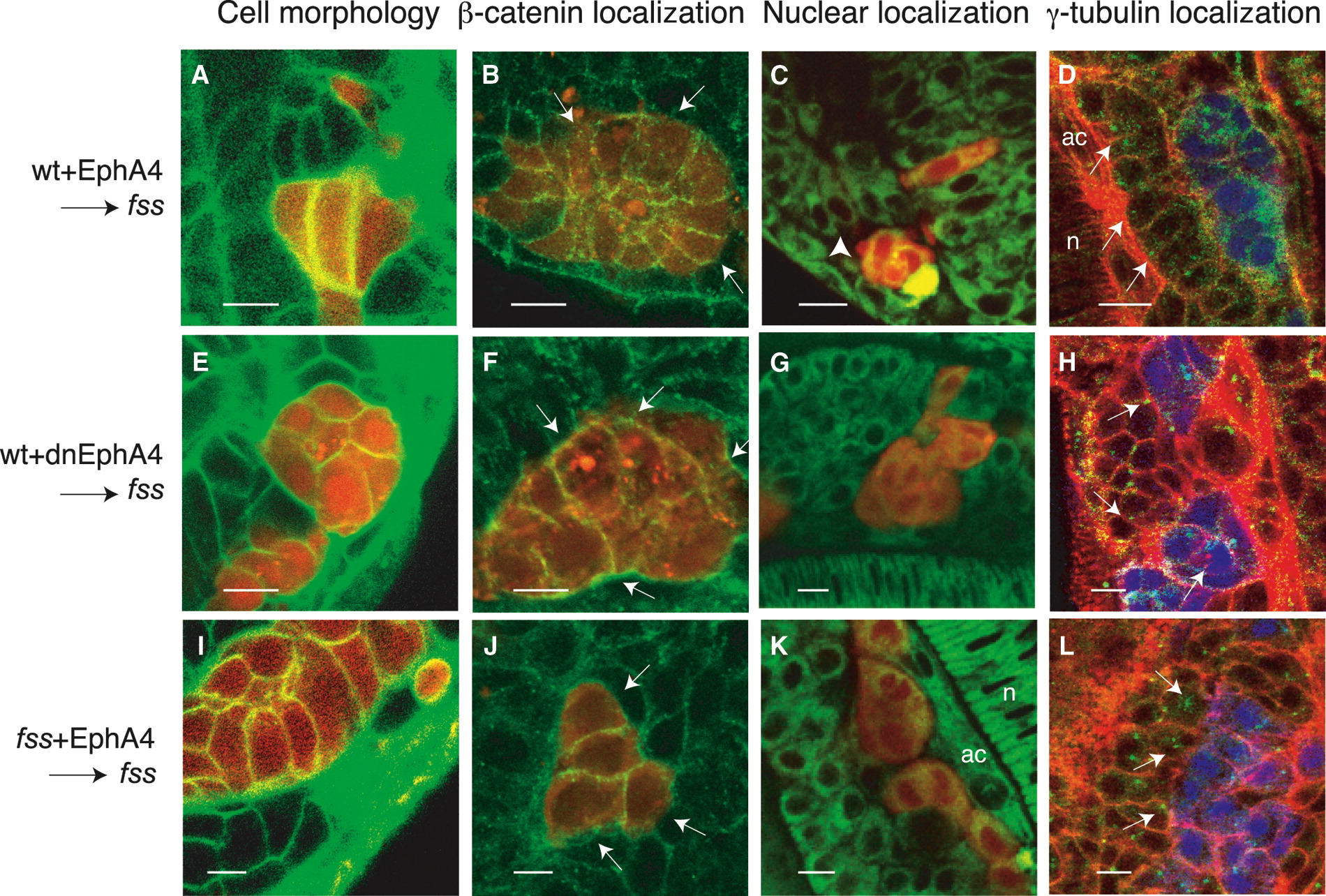
Fig. 5 Eph/Ephrin Signaling Rescues Epithelialization of Cells at Morphologically Distinct Boundaries
(A–L) (A, E, and I) Confocal images showing Bodipy ceramide-labeled somitic mesoderm of fss-/- host embryos (green) containing rhodamine dextran-labeled donor cells (red). (B, F, and J) Confocal images of β-catenin immunolocalization (green) in transplanted rhodamine dextran-labeled cells (red) in fss-/- host embryos. The arrows point to the basal surfaces of the transplanted cells at the interface at which morphologically distinct boundaries form (visible with DIC optics, not shown). In (B) and (J), β-catenin is reduced on the basal surfaces of these cells. (C, G, and K) Confocal images showing Bodipy 505-515-labeled somitic mesoderm of fss-/- host embryos (green) containing rhodamine dextran-labeled donor cells (red). The white arrowhead points to nuclei localized at the basal pole of host fss-/- cells, adjacent to the boundaries created between donor and host cells. (D, H, and L) Confocal images showing phalloidin-labeled somitic mesoderm of fss-/- host embryos (red) containing CFP-labeled donor cells (blue) and immunostained for γ-tubulin (green). The white arrows point to centrosomes. These are localized at the apical pole of host fss-/- cells adjacent to the boundary created between donor and host cells in (D) but are randomly positioned in (H) and (L). Donor cells are wild-type cells expressing (A–D) full-length or (E–H) truncated, dominant-negative EphA4 or fss-/- cells expressing (I–L) full-length EphA4. n, notochord; ac, adaxial cells. The scale bars represent 10 μm.