
|
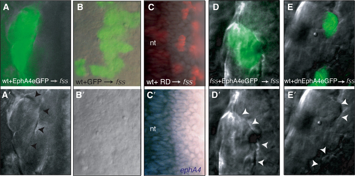
Fig. 3 Eph/Ephrin Signaling Restores Morphologically Distinct Boundaries in fss-/- Embryos
(A–E and A′–E′) (A–E) DIC and fluorescence overlays and (A′–E′) DIC images of the paraxial mesoderm of fss-/- hosts into which wild-type (wt) or fss-/- cells expressing various GFP-tagged reagents (green labeling in [A], [B], [D], and [E]) or containing rhodamine dextran (RD, red labeling in [C]) have been transplanted. Reagents are indicated at the bottom of the panels. (C′) ephA4 expression (blue) is absent from the transplanted cells. The arrowheads point to morphologically distinct boundaries formed at the interface between donor and host cells. nt, neural tube.