
|
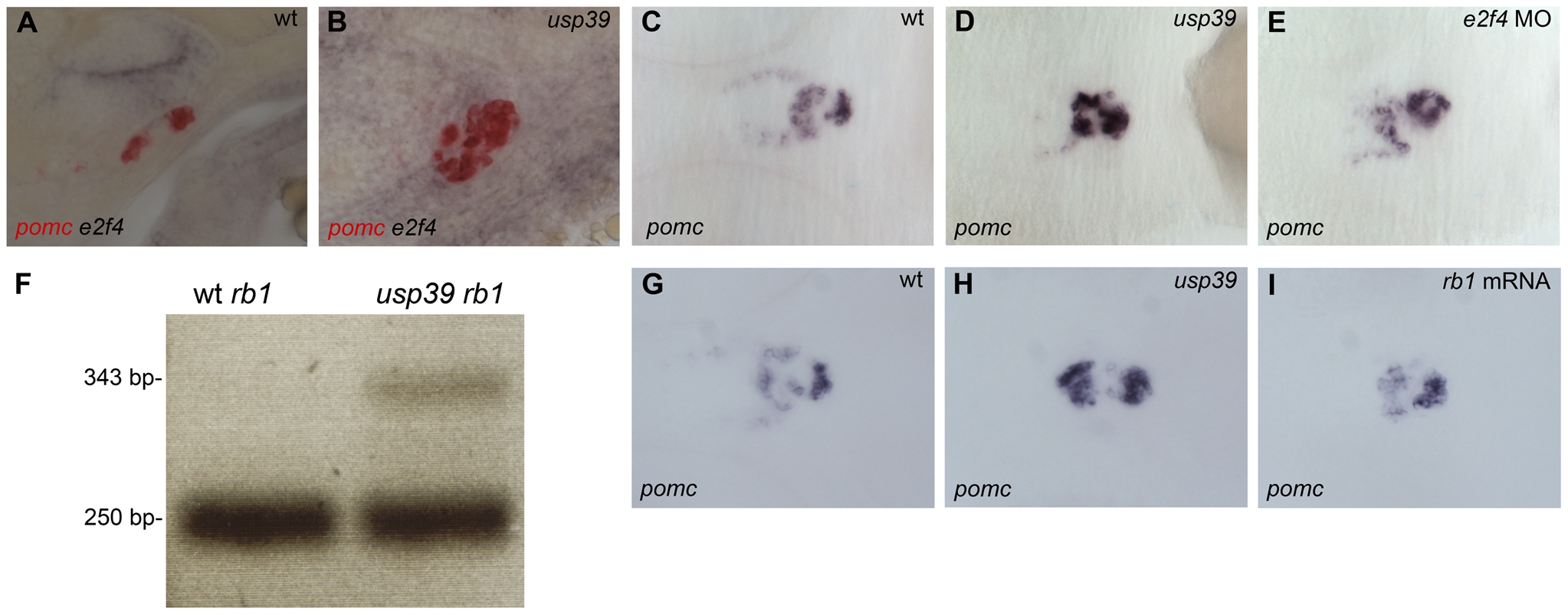
Fig. 6 Loss of usp39 leads to aberrant Rb1 mRNA splicing and increased pituitary e2f4 expression.
A,B: Whole-mount double in situ hybridization of pomc in red and e2f4 in purple at 48 hpf, lateral view. (A) Wild-type (wt) embryo. (B) Expression of e2f4 is higher and colocalizes with pomc expression in usp39 mutant embryos. C–E and G–I: Whole-mount in situ hybridization of pomc at 48 hpf, ventral view, anterior to left. (C) wt. (D) usp39 mutant. (E) e2f4-MO-injected usp39 mutant embryos showed partial pomc rescue similar to observed in wt embryonic pomc expression (C). F: PCR product with primers designed for region between exon 3 and exon 4 of rb1 in wt and usp39 mutant embryos. wt embryos only contain a 250 base pair (bp) PCR band, indicating that the intron was correctly spliced out. However, in the usp39 mutants there is an additional 343 bp band that contains the intron sequence. G–I: (G) wt. (H) usp39 mutant. (I) rb1 mRNA-injected usp39 mutants exhibit partial rescue of pomc expression similar to wt embryos (G).