
Image
|
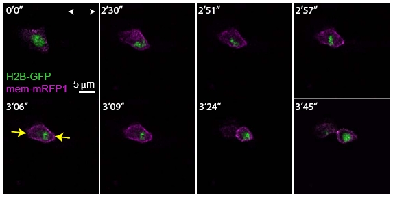
Figure Caption
Fig. S4
Rescue of Apicobasal Cell Division Orientation of a Single cdh2-/- Mutant Cell in a Wild- Type Environment
Selected time points from an in vivo imaging of a genetic mosaic in which a cdh2-/- donor derived cell (expressing H2B-GFP (green) and membrane bound mRFP1 (purple)) was transplanted into the presumptive posterior hindbrain of a WT host embryo. Double arrowhead indicates the apicobasal axis of the neuroepithelium, yellow arrows indicate the final orientation of cell division. Anterior is to the top. The times of the optical sections are given in min’s.
Acknowledgments
This image is the copyrighted work of the attributed author or publisher, and
ZFIN has permission only to display this image to its users.
Additional permissions should be obtained from the applicable author or publisher of the image.
Full text @ Curr. Biol.