
|
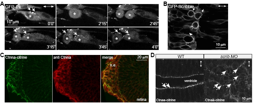
Fig. S3
GFP-Prickle Localizes to Cytoplasm, GFP-Scrib Shows Cortical Localization in WT Mitotic Neural Keel Progenitors, and Scrib Is Not Required for Maintenance of Subapical -Catenin Localization in the Mature Neural Tube Epithelium
(A) Optical sections from timelapse imaging (min’s“) revealing anterior polarization of GFP-Pk foci (arrows) in interphase and subsequent to cytokinesis (cells followed marked by stars), but absence of GFP-Pk foci in mitotic cells. Time of optical sections given in min’s“.
(B) Persistent cortical GFP-Scrib localization in WT neural keel progenitors with arrowhead noting a mitotic cell and arrows pointing to a cell undergoing cytokinesis. Dorsal horizontal view with anterior to the top in all panels. Double arrowheads indicate apicobasal axis.
(C) Ctnna-citrine fusion protein of Gt(ctnna-citrine) transgenic line colocalizes with the endogenous Ctnna protein in immunofluorescent stainings. Single confocal images taken in the region of nasal epithelium (n.e.) and neuroepithelium of the retina.
(D) Ctnna-citrine localization (arrows) in the mature neural tube reveals residual Ctnna-citrine foci in the disorganized hindbrain epithelium of scrib morphants. Double arrowheads indicate apicobasal axis. Anterior-posterior axis is horizontal.