
|
Fig. S2
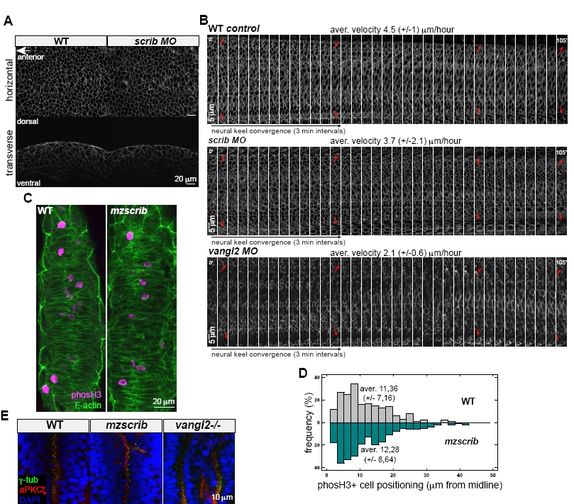
Scrib Is Not Essential for Proper Velocity of Hindbrain Neural Keel Cell Convergence, and Scrib Shows Different Effects on Zebrafish Hindbrain Organization from Vangl2
(A) Single optical slices of WT and scrib morphant hindbrain neural plate architecture in dorsal horizontal view (upper row) and transverse view (lower row) with membrane-bound GFP at 1-2 somites (10.5 hpf).
(B) Hindbrain neural keel convergence analyzed by multi-photon imaging of membrane-bound GFP in dorsal horizontal view starting at 4 somites (11.3 hpf). An image was acquired every 3 minutes for 105 minutes, ending at the 8 somite stage. Pictures were taken at all z-levels of the neural keel and the broadest keel positions were used for the analysis. Note abnormal convergence in the vangl2 morphant and only slightly slower convergence in scrib morphant compared to WT. Arrows indicate the same cells at different time points. Anterior is to the left in all panels. Average neural keel convergence velocity in the posterior hindbrain noted on top of each panel (n = 2 for each genotype).
(C) Medial positioning of mitotically dividing cells marked with anti phosphohistone H3 (Ser10) medially along the midline in the posterior hindbrain neural keel of WT as well as in mzscrib mutants at 14 hpf. Images present maximal intensity projections of 4.8 μm confocal optical sections in dorsal horizontal view, anterior is to the top.
(D) Quantification of the positioning of phosphohistone H3 positive mitotic cells relative to the midline in confocal sections of the dorsal 30 m of the neural keel plotted in μm on the horizontal axis. The y-axis displays distribution frequency. Mitotic cells were positioned at an average distance of 11.36 ± 7.16 μm (n = 196 cells, 9 embryos) in the WT (grey) and 12.28 ± 8.64 μm (n = 238 cells, 9 embryos) in the mzscrib mutants (green). The differences between the two populations were not significant (p > 0.2, t-test).
(E) Organization of hindbrain neural progenitors in horizontal dorsal view showing the abnormally branched midline in mzscrib compared to the double midlines of vangl2-/- mutants. aPKCζ (red), γ-tubulin (green) and DAPI (blue). Anterior to the top in all panels.