
|
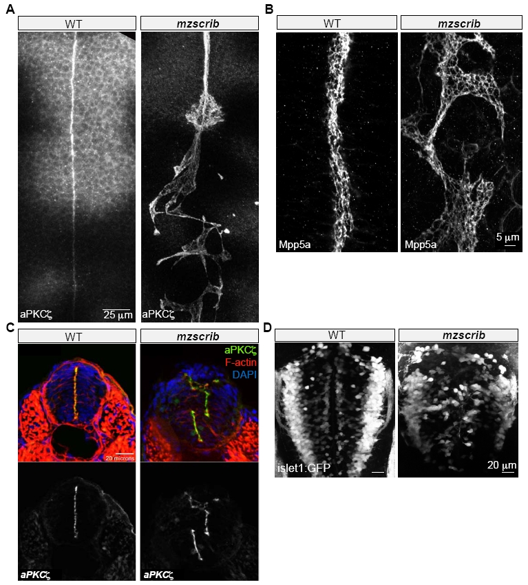
Fig. S1
Requirement of Scrib in Zebrafish Posterior Hindbrain Organization
(A) Posterior hindbrain structure in mzscrib mutants as analyzed by aPKCζ staining. Labeled apical cell membranes are aligned at the midline of WT embryos and misaligned in mzscrib mutants.
(B) Alignment of the subapically localized tight junctional component Mpp5a at the midline in WT and aberrant alignment in mzscrib mutants. These are the same embryos as shown in Figure 1D, imaged in a different channel.
(C) Abnormal organization of the neural tube lumen in mzscrib compared to WT in transverse sections at posterior hindbrain level as analyzed by aPKCζ green), F-actin (red) and DAPI (blue) staining. Dorsal is to the top.
(D) mzscrib mutants display aberrant vagus motor neuron positioning visualized in tg(isl1:GFP) transgenic zebrafish. Dorsal view. Anterior is to the top in A,B,D.