
|
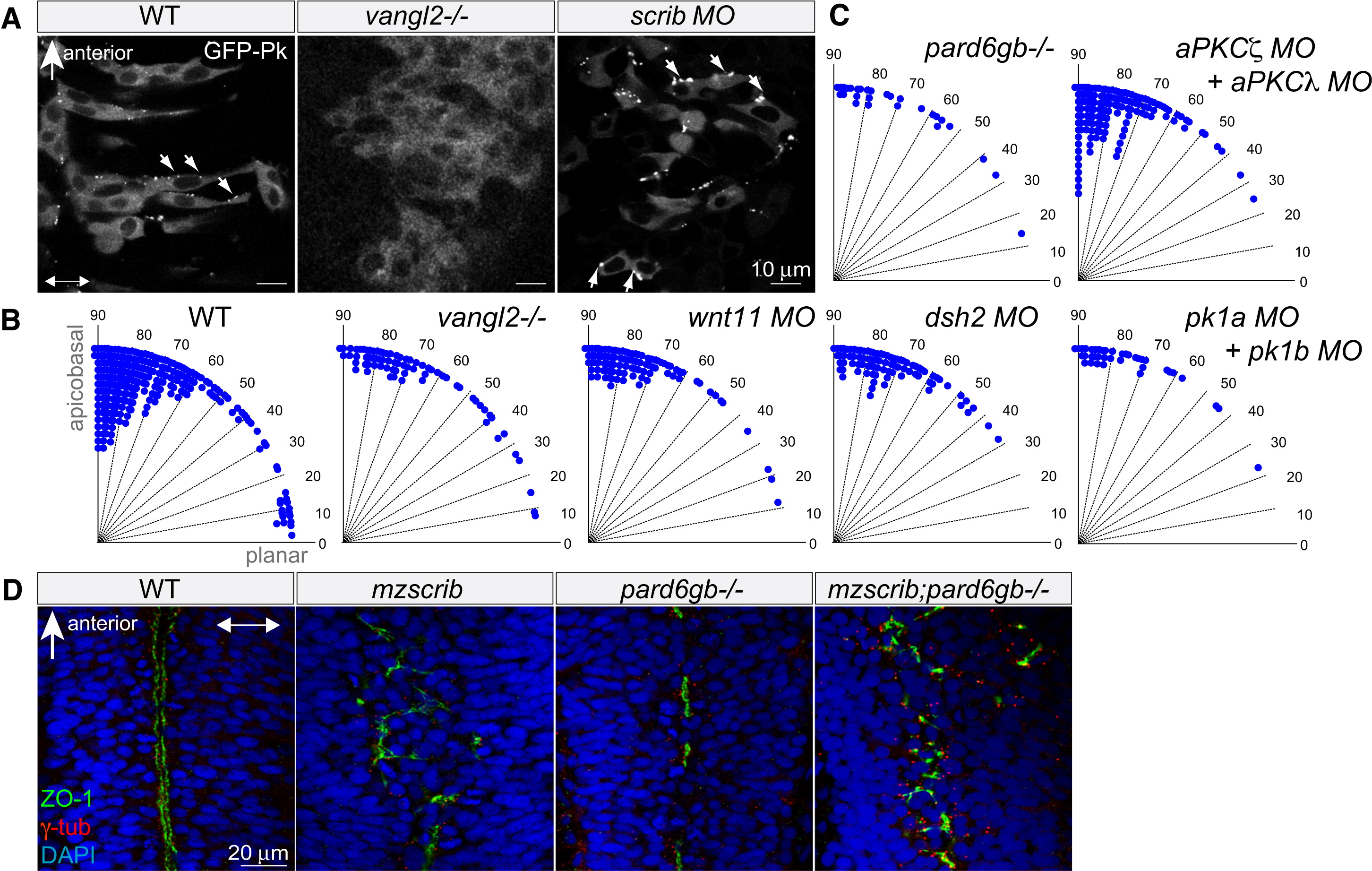
Fig. 2 Neither Planar Cell Polarity Components nor the Par Complex Are Required for Proper Spindle Orientation in the Neural Keel
(A) GFP-Prickle (GFP-Pk)-positive foci (arrows) in posterior hindbrain neural keel progenitors of WT, vangl2-/- mutant, and scrib morphant embryos. In (A) and (D), anterior is to the top and two-way arrows indicate the apicobasal axis.
(B and C) Quantification of anaphase orientation without functional planar cell polarity and Par complex components. The distributions of the various mutant or morphant conditions shown are not significantly different from WT (n = 269): vangl2-/- mutant (n = 72, χ2 = 5.1, 4 df; p = 0.28), wnt11 morphant (n = 74, χ2 = 2.6, 3 df; p = 0.41), dsh2 morphant (n = 86, χ2 = 9.6, 4 df; p = 0.05), pk1a + pk1b morphant (n = 38, χ2 = 2.6, 3 df; p = 0.46), pard6gb-/- mutant (n = 28, χ2 = 2.7, 3 df; p = 0.43); the distribution of mitotic angles in aPKCζ + aPKCλ double morphants is slightly more biased toward apicobasal than WT (n = 139, χ2 = 14.4, 4 df; p = 0.006).
(D) The branched, disorganized neural tube lumen of mzscrib and mzscrib;pard6gb-/- double mutants. Dorsal optical sections at 18 hpf immunostained with γ-tubulin (red), ZO-1 (green), and DAPI (blue) are shown.
See also Figure S2.