
|
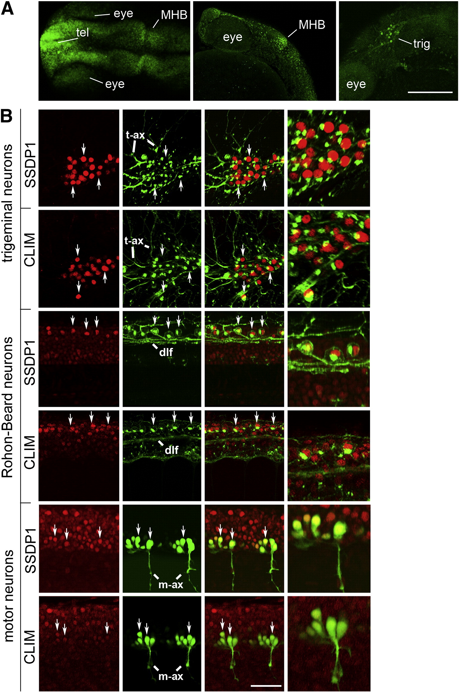
Fig. 2 SSDP1 protein expression corresponds to in situ hybridization patterns and SSDP1 is co-expressed with CLIM protein in neuronal cell types. A: Dorsal (left) and lateral (middle, right) views of the head labeled with SSDP1 antibodies at 24 hpf are shown. Strong immunoreactivity is detectable in the telencephalon (tel), in the MHB and in the trigeminal ganglion (trig). The right panel is a selective z-stack of the epidermal region. Scale bar: 200 μm. B: Neuronal localization of SSDP1 and CLIM immunoreactivity (red) is revealed by co-labeling with an HNK-1 antibody (trigeminal and Rohon–Beard neurons in green) or by transgenic GFP expression in HB9:GFP transgenic embryos (motor neurons in green) at 24 hpf. The right outer column gives a higher magnification of double-labeled neurons. HNK-1 is strongly labeled on axonal membranes and Golgi apparatus next to nuclear labeling of SSDP1 and CLIM in trigeminal and Rohon–Beard neuron. In GFP expressing motor neurons, nuclear labeling is found inside the somata. Some double-labeled neurons are indicated by arrows. Trigeminal (t-ax) and motor axons (m-ax) as well as the dorsal longitudinal fascicle (dlf) are indicated for orientation. Scale bar: 50 μm for low and 100 μm for high magnification.
Reprinted from Developmental Biology, 349(2), Zhong, Z., Ma, H., Taniguchi-Ishigaki, N., Nagarajan, L., Becker, C.G., Bach, I., and Becker, T., SSDP cofactors regulate neural patterning and differentiation of specific axonal projections, 213-224, Copyright (2011) with permission from Elsevier. Full text @ Dev. Biol.