
Image
|
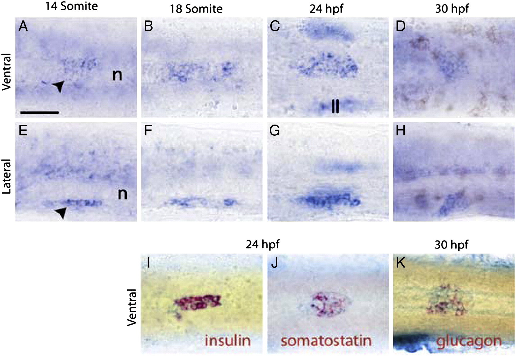
Figure Caption
Fig. 1 Expression of dachb in the zebrafish pancreas. Whole-mount in situ hybridization was performed for dachb (A–K, blue) and insulin (I, red), somatostatin (J, red), and glucagon (K, red) at the stages of zebrafish development shown. Ventral views (A–D;I–K), and lateral views (E–H) are shown with anterior to the left. On all the panels the yolk was manually removed from the embryos. The notochord (n) and the lateral line (ll) have been indicated in panels A, E and C. Black arrowheads: dachb-expressing cell in the pancreatic region. Scale bar, 50 μm.
Figure Data
Acknowledgments
This image is the copyrighted work of the attributed author or publisher, and
ZFIN has permission only to display this image to its users.
Additional permissions should be obtained from the applicable author or publisher of the image.
Reprinted from Developmental Biology, 348(2), Kalousova, A., Mavropoulos, A., Adams, B.A., Nekrep, N., Li, Z., Krauss, S., Stainier, D.Y., and German, M.S., Dachshund homologues play a conserved role in islet cell development, 143-152, Copyright (2010) with permission from Elsevier. Full text @ Dev. Biol.