
Image
|
Figure Caption
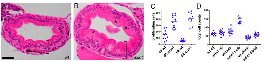
Fig. 3 Up-regulation of Wnt signaling causes intestinal hyperplasia. (A) H&E-stained section of an 8-dpf WT (wt) intestinal bulb shows an orderly array of cells in the intestinal epithelium. (B) An 8-dpf axin1 mutant intestine is thicker (bracket) and has more disorganized epithelial cells (*). (C) At both 6 and 8 dpf, axin1 mutants have significantly more BrdU-labeled cells within the intestinal epithelium than wt (P < 0.0001). (D) At 8 dpf, axin1 mutant intestines have significantly more epithelial cells in the bulb and distal intestine vs. wt (P < 0.0001). (Scale bar: A and B, 25 μM.) 4
Figure Data
Acknowledgments
This image is the copyrighted work of the attributed author or publisher, and
ZFIN has permission only to display this image to its users.
Additional permissions should be obtained from the applicable author or publisher of the image.
Full text @ Proc. Natl. Acad. Sci. USA