
|
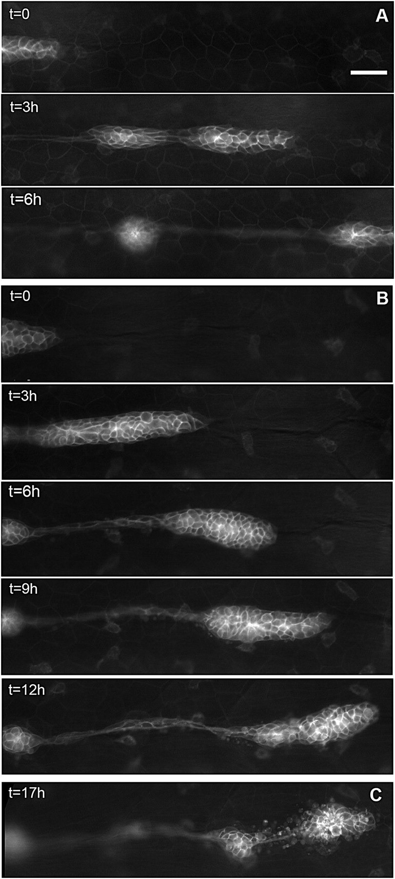
Fig. 2 Migration of the PLL primordium is altered in esr1-MO embryos. (A) Three frames of a time-lapse movie of primordium migration in a cldnb:gfp embryo at 35 (t = 0), 38, and 41 hpf, respectively. At 41 hpf, the primordium has just migrated out of the field. (B) Five frames of a time-lapse movie of the primordium in an esr1-MO1 injected embryo at 35 (t = 0), 38, 41, 44, and 48 hpf, respectively. Migration is two to three times slower than in untreated cldnb:gfp embryos and eventually comes to a halt around 45 hpf. (C) In the same embryo, the immobile primordium has fragmented into two neuromasts at 51 hpf. (Scale bar in A: 50 μm.)