
|
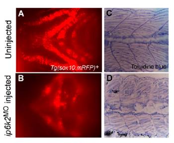
Fig. S4 ip6k2 knockdown perturbs craniofacial development and somite structures. (A and B) Ventral view of the head region showing cartilage fluorescence of 3-d-old Tg(sox10(7.2):mrfp) embryo (A) and embryo injected with ip6k2ATGMO (B). Reduced mRFP expression in the ip6k2ATGMO embryos reflects a lack of pharyngeal arches. (C and D) Toluidine blue staining of the trunk region of 5-d-old uninjected (C) and ip6k2ATGMO-injected (D) embryo. The embryos were fixed and embedded in plastic resin, sectioned, and stained with toluidine blue. Sagittal sections through the midtrunk region were shown. The somites in the ip6k2ATGMO embryo were defective in shape and muscle fiber organization.