
|
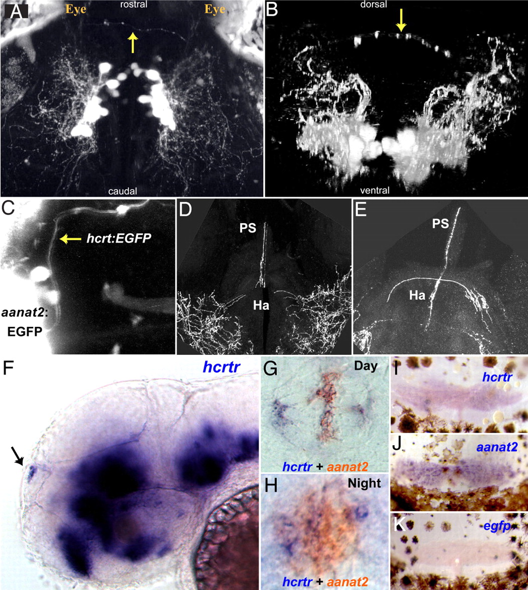
Fig. 4
The HCRT-pineal gland circuit. (A and B) Dorsal and frontal views of the brain of a 7 dpf hcrt:EGFP transgenic larva imaged by two-photon microscopy. HCRT axons (arrows) projecting toward the pineal gland are observed. (C) A dorsal image of 6 dpf transgenic larva carrying two transgenes; an EGFP reporter driven by hcrt (hcrt:EGFP) and the pineal-specific aanat2 (aanat2:EGFP) promoters, demonstrate direct axon projection (arrow) to the pineal gland. (D and E) Close-ups of two adjacent transversal hcrt:EGFP adult brain sections showing HCRT projections to the habenula and the pineal gland stalk. (F) Lateral and (G and H) dorsal views of whole-mount in situ hybridization of 2-dpf embryos. (F) hcrtr mRNA is expressed in several regions of the brain (16) including the pineal gland (arrow). Double ISH experiment with aanat2 demonstrates that hcrtr is expressed in the pineal gland during the day (G) and the night (H). Similarly, in adult animals, hcrtr is expressed in the pineal gland (I). aanat2 (J) and egfp (K) probes were used as positive and negative controls, respectively. Adult pineal glands (I–K) were removed with the upper skull and skin hence presence of brown melanophores cells in the preparations.