
|
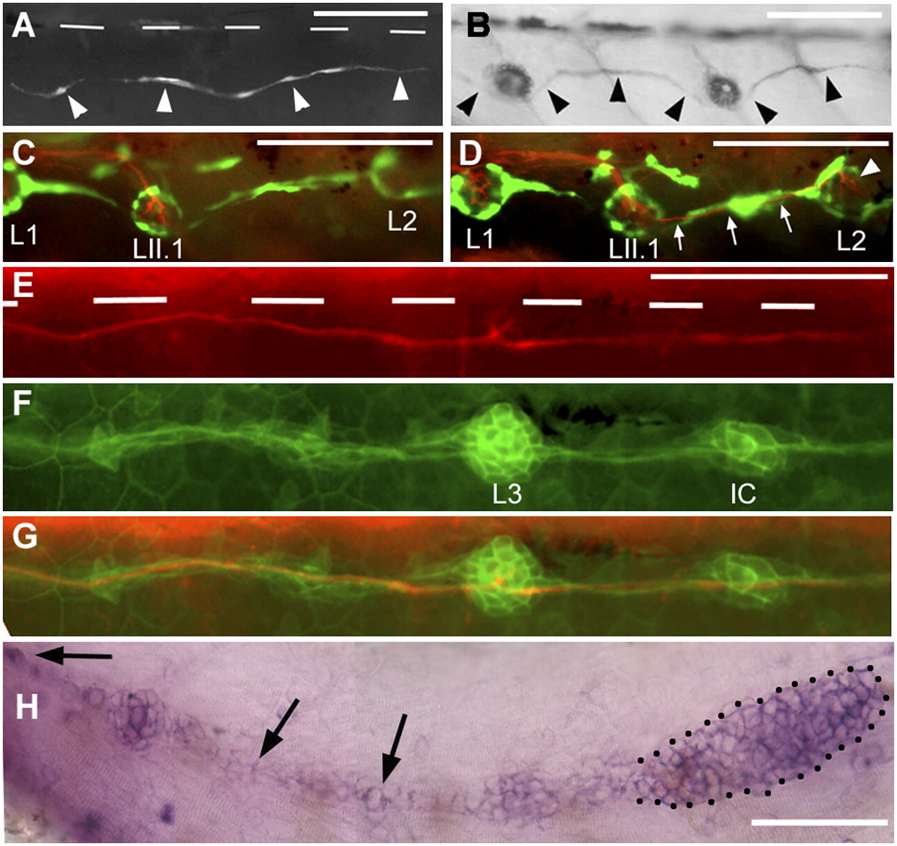
Fig. 5
Regenerating axons can be guided by interneuromast cells. (A) After ablation of glial cells in the foxd3:gfp line, new glial cells extend posteriorly (arrowheads). (B) Their path resembles that of interneuromast cells (arrowheads) as visualized by alkaline phosphatase labeling. (C and D) After glial ablation between L1 and L2 in 3-dpf Huc:kaede, Et20:gfp fish, axons regrow along the myoseptum up to LII.1 (C) and follow the interneuromast cells (arrows) up to L2 (arrowhead in D). (E) Regenerating axons in a 54-hpf Huc:kaede, cldnb:gfp fish where the nerve had been cut at 30 hpf. White bars indicate the position of the horizontal myoseptum. (F) Trail of interneuromast cells in the same embryo; (G) merge. (H) Expression of gdnf in the primordium (dotted outline) and in the interneuromast cells (arrows) as revealed by in situ hybridization. (Scale bar, 100 mm.) A and H were assembled from two consecutive levels in Z-stacks.