
|
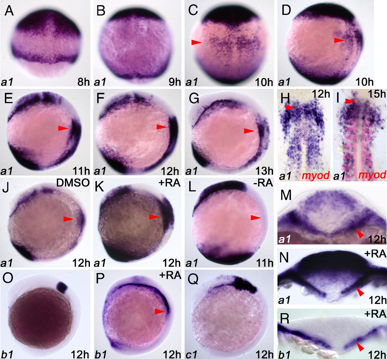
Fig. 1 Expression of cyp26 genes in late zebrafish gastrulae. (A–G) Expression of cyp26a1 from 8 to 13 hpf. Shown are whole mounts, animal pole up, dorsal views (A–C) and lateral views (D–G). Arrowheads indicate endodermal expression. (H and I) Double in situ hybridizations with cyp26a1 (purple) and myod (red) to show paraxial mesoderm. Embryos are deyolked and flat-mounted, and anterior is to top of page. Arrowheads indicate anterior limit of myod expression. (J) cyp26a1 expression in DMSO carrier-treated embryo at 12 hpf, whole-mount, lateral view. (K) cyp26a1 expression in embryo treated with 0.1 μM RA from 5.25 hpf; note up-regulation of expression. (L) cyp26a1 expression in 10 μM 4-(diethylamino)benzaldehyde (DEAB)-treated (RA-deficient) embryo from 5.25 hpf; note absence of anterior trunk expression. (M) Transverse optical section through anterior trunk at 12 hpf. (N) Transverse section through anterior trunk of RA-treated embryo as in K; note up-regulation of endodermal cyp26a1. (O) cyp26b1 expression at 12 hpf, whole-mount, lateral view. (P) cyp26b1 expression in embryo treated with 0.1 μM RA from 5.25 hpf. (Q) cyp26c1 expression at 12 hpf, whole-mount, lateral view. (R) Transverse section through anterior trunk, embryo treated with 0.1 μM RA from 5.25 hpf; note up-regulation of endodermal cyp26b1. Arrowhead indicates anterior trunk, and for M, N, and R arrowheads indicates anterior trunk endoderm.