
|
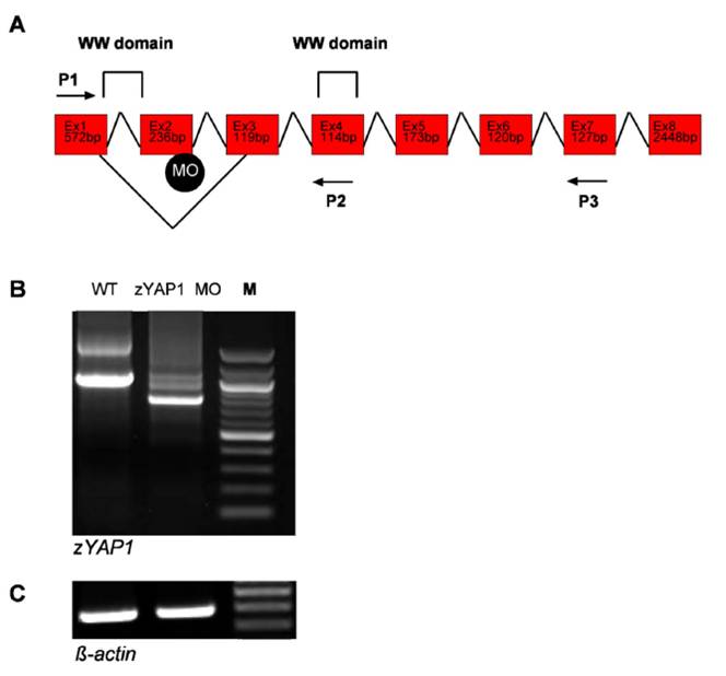
Fig. S6 Defective splicing within theWWdomain of zebrafish zYAP1. (A) Schematic representation of zYAP1 and the MO-induced splicing defect within the first WW domain. The WW domain is encoded by Exon1 (Ex1) and Exon2 (Ex2). Position of the MO targeted against the donor splice site is indicated. Two sets of PCR primer combinations, P1/P3 and P1/P2, were used to detect the splice defect. (B) Analysis of the splicing defect induced by the zYAP1 MO. RT-PCR of YAP1 at 55 h.p.f generated a 1031-bp fragment in wild-type embryos (Lane WT), and a 777-bp fragment in embryos injected with zYAP1 MO (Lane zYAP1 MO). M, 100-bp marker. The defective splicing resulted in preterminal stop of the protein. (C) RT-PCR of β-actin mRNA was performed to control for equal amounts of RNA in each sample.