
|
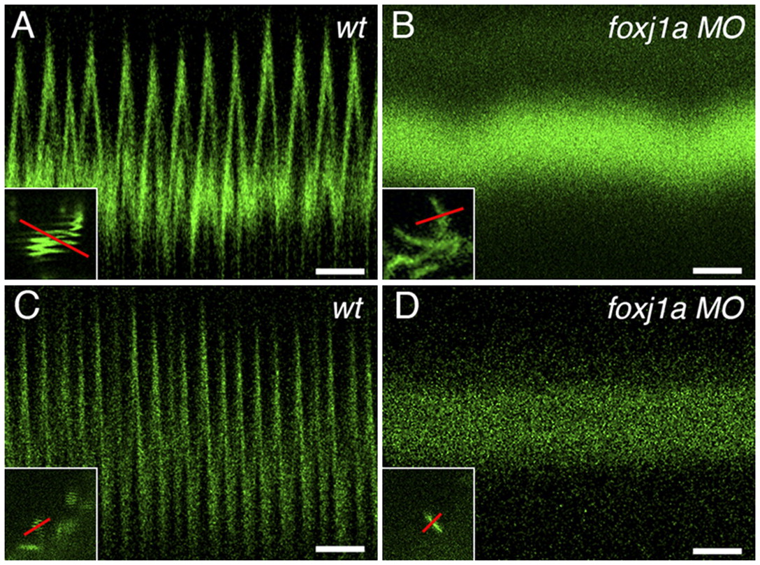
Fig. 3
foxj1a is required for cilia motility. (A) Control scorpion-eGFP-labeled spinal canal cilia in 24-hpf embryos beat rapidly as seen in high-speed line scanning over time (16.9 beats/s in this example; total time, 768 ms. (Scale bar, 100 ms.) (A, Inset) Single cilia imaged in slow-frame scan appears as squiggled line pattern; red line in Inset indicates line acquired in high-speed line scan. (B) High-speed line scan of foxj1a 24-hpf morphant spinal cord cilia shows no motility (total time, 768 ms). (Scale bar, 100 ms.) (B, Inset) Single cilia imaged in slow frame scan shows no movement; red line in inset indicates line acquired in high-speed line scan. (C) Control Kupffer′s vesicle cilia (12-somite stage) beat rapidly, as seen in high-speed line scanning over time (27 beats/s in this example; total time, 768 ms). (Scale bar, 100 ms.) (C, Inset) Single cilia at edge of Kupffer′s vesicle imaged in slow frame scan appears as squiggled line pattern; red line in Inset indicates line acquired in high-speed line scan. (D) High-speed line scan of foxj1a morphant Kupffer′s cilia (12-somite stage) shows no motility (total time, 768 ms). (Scale bar, 100 ms.) (D, Inset) Single cilia imaged in slow frame scan shows no movement; red line in inset indicates line acquired in high-speed line scan.