
Image
|
Figure Caption
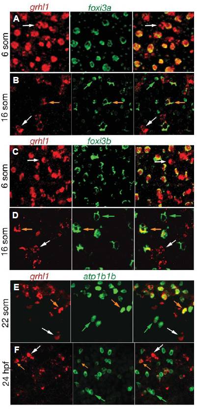
Fig. 3 grhl1 is transiently expressed in ionocytes. All panels show double fluorescent in situ hybridisations with probes indicated above the panels. The right picture of each panel shows the merge of the two left pictures. Stages are indicated at the left. All panels show cells of wild-type embryos over the yolk sac (5 -16 som) or over the anterior half of the trunk (22 som - 24 hpf). White arrows mark cells expressing only grhl1, green arrows mark cells only expressing foxi3a (B), foxi3b (D), or atp1b1b (F), and orange arrows mark cells co-expressing the two indicated genes.
Figure Data
Acknowledgments
This image is the copyrighted work of the attributed author or publisher, and
ZFIN has permission only to display this image to its users.
Additional permissions should be obtained from the applicable author or publisher of the image.
Full text @ Int. J. Dev. Biol.