
Image
|
Figure Caption
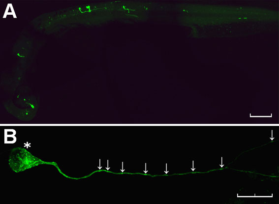
Fig. S2 Mosaic expression of NBT:EB3-GFP in a zebrafish embryo. (A) Low-power view of a fixed embryo shows GFP fluorescence in a scattered population of neurons distributed randomly throughout the brain and spinal cord (anterior to left, dorsal top). (B) High-power view of a single EB3-GFP-expressing neuron in the CNS of a fixed zebrafish embryo reveals discrete labelling of puncta in the cell body and throughout the length of the axon (arrows). Scale bars: 100 μm (A) and 20 μm (B).
Acknowledgments
This image is the copyrighted work of the attributed author or publisher, and
ZFIN has permission only to display this image to its users.
Additional permissions should be obtained from the applicable author or publisher of the image.
Full text @ Dis. Model. Mech.