
Image
|
Figure Caption
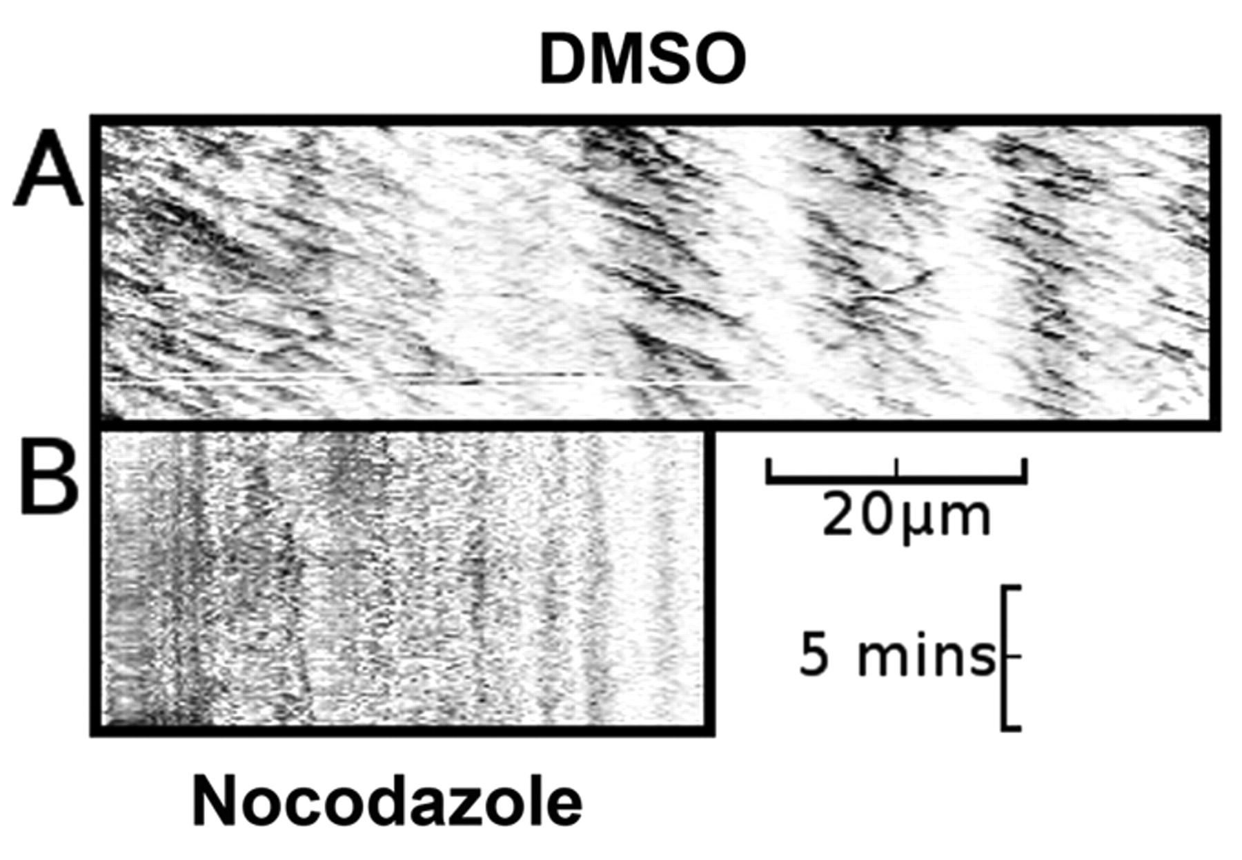
Fig. 5 Nocodazole-treated embryos lack both EB3-GFP puncta and axonal movement of EB3-GFP. (A) Kymograph of an axon made from a 10-minute time-lapse recording of a neuron from a DMSO-vehicle-treated embryo, demonstrating predominantly anterograde movement of discrete puncta of EB3-GFP fluorescence (n=16). (B) Kymograph of an axon made from a 10-minute time-lapse recording of a neuron from a nocodazole-treated embryo, demonstrating absence of axonal movement of EB3-GFP fluorescence (n=10). Horizontal scale bar: 20 μm, vertical scale bar: 5 minutes. See supplementary material Movies 7 and 8.
Acknowledgments
This image is the copyrighted work of the attributed author or publisher, and
ZFIN has permission only to display this image to its users.
Additional permissions should be obtained from the applicable author or publisher of the image.
Full text @ Dis. Model. Mech.