
|
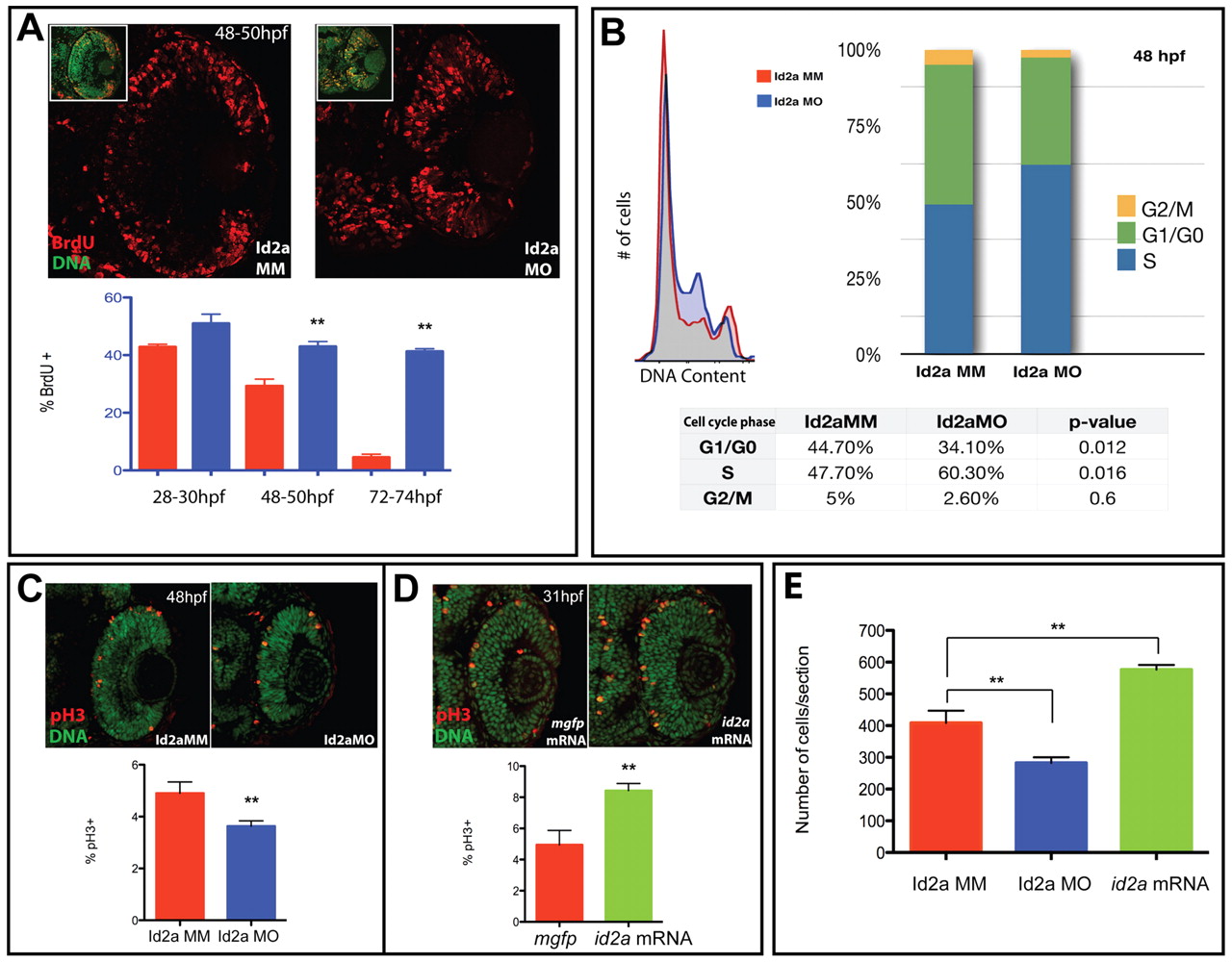
Fig. 5 Id2a is required for S-phase progression and is sufficient to enhance retinoblast mitotic activity. (A) BrdU exposures from 28-30, 48-50 and 72-74 hpf reveal an increased proportion and mislocalization of S-phase cells in Id2a-MO retinas compared with Id2a-MM retinas. n=4 retinas; **, P<0.05. Insets are examples of SYTOX Green/BrdU overlays used for cell counts. (B) FACS DNA-content analysis of Id2a-MM and Id2a-MO retinas indicates that Id2a-MO retinal cells contain an increased percentage of retinoblasts in S phase, concomitant with decreases in G0/G1 and G2/M (data averaged from three independent experiments). (C) pH3 localization in Id2a-MM and Id2a-MO retinas at 48 hpf. n=5 retinas; **, P<0.05. (D) pH3 in mgfp mRNA-injected and id2a-overexpressing retinas at 31 hpf. Id2a-MO retinas contain a lower percentage of mitotic cells and id2a-overexpressing retinas contain a higher percentage than controls. n=5 retinas; **, P<0.05. (E) Average cell numbers in Id2a-MM, Id2a-MO and id2a-overexpressing retinas. n=4 retinas; **, P<0.05. Error bars indicate ± s.e.m.