
|
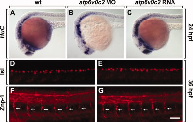
Fig. 5 ATP6V0C2 function is dispensable for neurogenesis. All panels show lateral views of whole embryos, anterior to the left. A–C: Wild-type (A), atp6v0c2 MO-injected (B), and atp6v0c2 RNA-injected (C) embryos analyzed by HuC RNA in situ hybridization. D, E: Labeling with an anti-Isl antibody to stain for sensory neurons in the dorsal spinal cord of wild-type (D) and atp6v0c2 MO-injected (E) embryos. F, G: Labeling with an anti-Znp-1 antibody to stain for motor axon fibers in the peripheral nervous system of wild-type (F) and atp6v0c2 MO-injected (G) embryos. Arrows indicate motor axon fibers. Scale bar: A–F, 100 μm; G–J, 40 μm.