
|
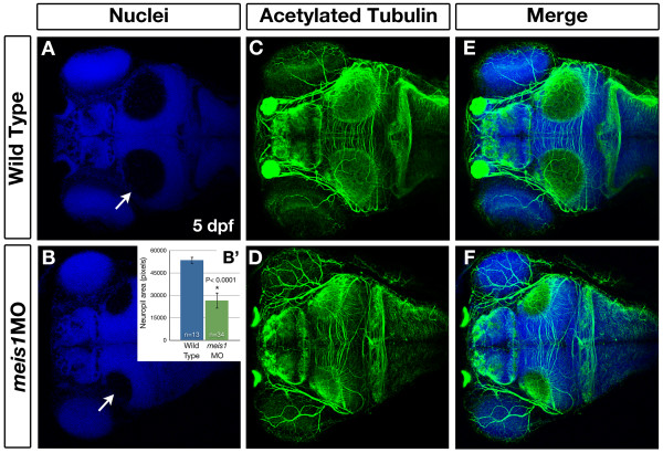
Fig. 9 Meis1-depleted embryos have smaller tectal neuropil at 5 dpf. (A-F) Whole mount immunohistochemistry using anti-acetylated tubulin (axons - green) and Hoechst 33258 (nuclei - blue) to compare the size of the tectal neuropil in 5-dpf wild type (A, C, E) and meis1 morphant (meis1MO) (B, D, F) embryos. White arrows in (A, B) indicate the tectal neuropil. (B′) The area (in pixels) of the neuropil from wild type and meis1 morphant embryos was measured using ImageJ. The n values represent individual neuropil regions. The error bars show plus/minus one standard deviation. The asterisk indicates a statistically significant reduction the size of morphant neuropil as determined by an unpaired, two-tailed t-test.