
|
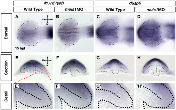
Fig. 7 Retinal Fgf signalling is upregulated in Meis1-depleted embryos. (A-D) mRNA in situ hybridization (ISH) for the Fgf-responsive genes il17rd/sef (A, B) and dusp6 (C, D) in wild-type (A, C) and meis1 morphant (meis1MO) (B, D) embryos. Dotted lines outline the optic vesicle. The vertical dotted line in (A) indicates the estimated position of the transverse sections in (E-H). Views are dorsal with anterior left. (E-H) Transverse sections through the eyes of 15-hpf wild-type and meis1 morphant embryos stained for il17rd and dusp6. (E′-H′) Detailed views of the corresponding sections in (E-H). The region of interest is indicated by the red dashed-line box. Dotted lines outline the optic vesicles. All transverse sections are oriented with dorsal up. Legend for retinal axial position: D, dorsal; V, ventral; N, nasal; T, temporal; L, lateral; M, medial; A, anterior; P, posterior.