
Fig. S2
|
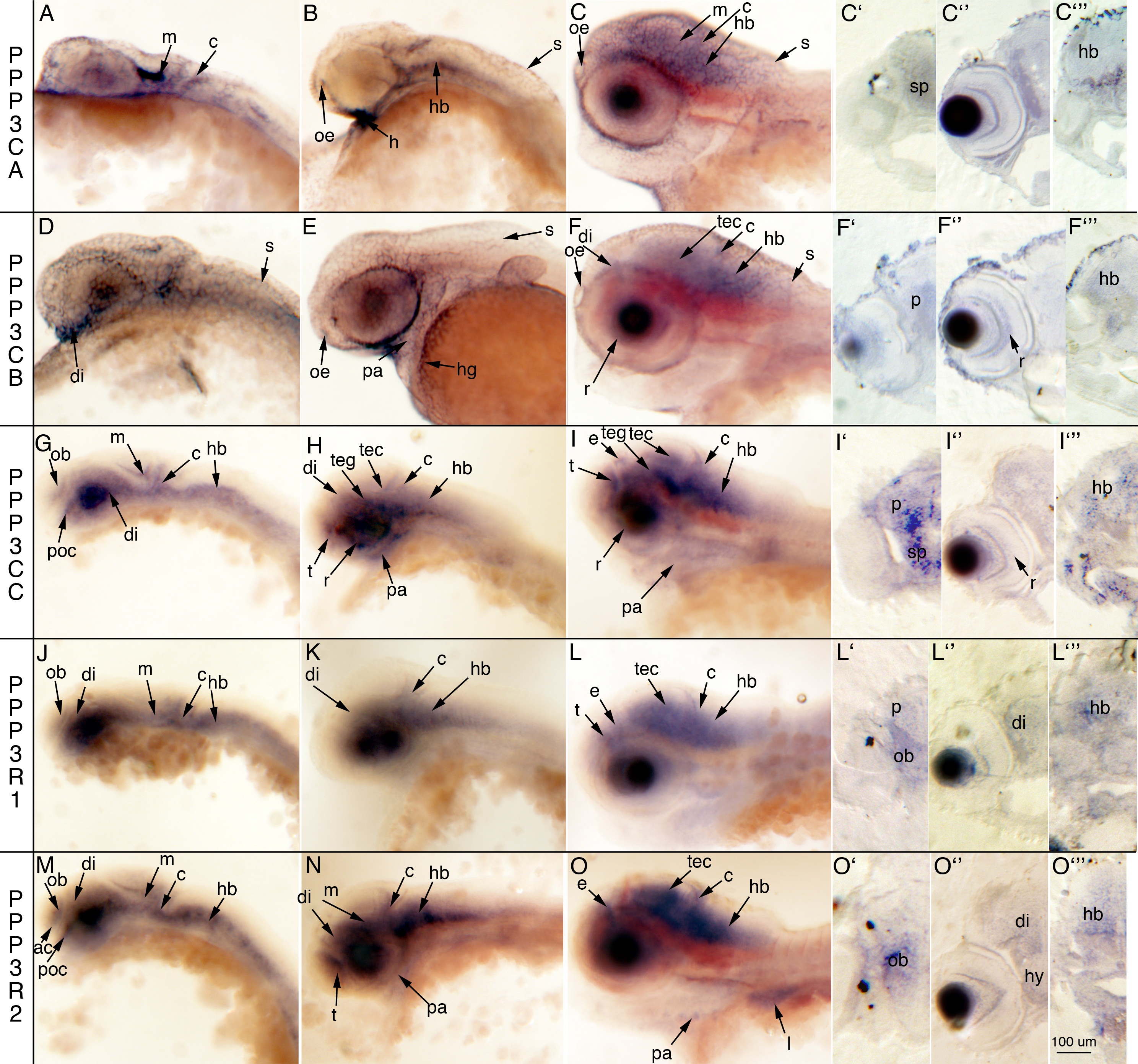
Fig. S2 Calcineurin subunits are expressed in unique temporal and spatial patterns. All calcineurin subunits are expressed in the nervous system during development as determined by whole embryo in situ hybridization (ISH). A–O: Lateral views of the head at 24 hpf (A,D,G,J,M), 48 hpf (B,E,H,K,N), and 72 hpf (C,F,I,L,O) and coronal sections of stained embryos at 72 hours postfertilization (hpf; C2–C23 F2–F23 I2–I23 L2–L23 O2–O23). ac, anterior commissure; c, cerebellum; di, diencephalon; e, epiphysis; h, heart; hb, hindbrain; hg, hatching gland; hy, hypothalamus; l, liver; m, midbrain; ob, olfactory bulb; oe, olfactory epithelium; p, pallium; pa, pharyngeal arches; poc, post optic commissure; r, retina; s, skin; sp, subpallium; t, telencephalon; tec, tectum; teg, tegmentum. Some embryos have been manually deyolked.