
|
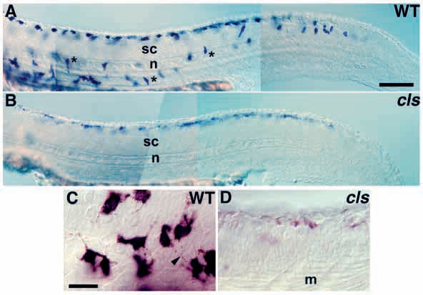
Fig. 1
Melanoblasts are abnormal in cls- embryos.
(A) Lateral view of wild-type embryo labeled for dct transcripts at 27 hpf reveals abundant melanoblasts along trunk and tail, with many along medial migration pathway (*). (B) cls- embryos have fewer melanoblasts, all dorsal to spinal cord (sc). (C) At higher magnification, wild-type melanophores show intense cytoplasmic dct-labeling (purple) and melanosomes (brown granules, arrowhead) in processes. (D) cls- melanophores show only faint dct labeling. In these and all subsequent lateral views, anterior is to the left and dorsal to the top. At both 24 and 27 hpf cls- embryos show only approximately one third the number of melanoblasts of wild types (data not shown). m, muscle fibres; n, notochord. Scale bar, 250 μm (A,B) and 50 μm (C,D).