
Fig. 1
- ID
- ZDB-IMAGE-100910-1
- Genes
- Antibodies
- Publication
- Coutelle et al., 2001 - Hedgehog signalling is required for maintenance of myf5 and myoD expression and timely terminal differentiation in zebrafish adaxial myogenesis
- All Figures
- Figures for Coutelle et al., 2001
|
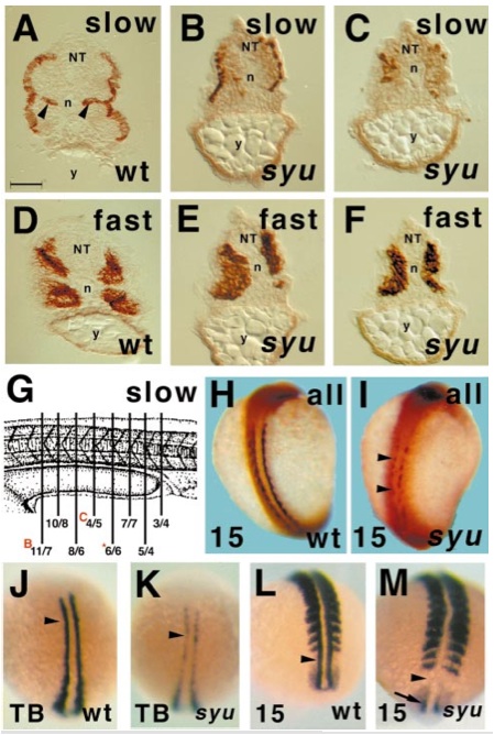
Fig. 1 Sonic you zebrafish mutants have deficits in slow muscle. Transverse sections show that somitic muscle of sibling wild type or heterozygote (wt, A, D) and homozygous mutant sonic you (syu, B, C, E, F, I) 24-hpf zebrafish embryos contain superficial slow myosin-immunoreactive muscle fibres (A–C, I) and medial fast myosin-immunoreactive muscle fibres (D–F). Note that syu embryos entirely lack the medial slow muscle pioneer fibres observed in the nonmutant siblings (compare B, C with arrowheads in A) and show a progressive loss of lateral slow muscle fibres from rostral regions (not shown), through rostral yolk extension regions (A, B, somite ~15) to caudal yolk extension (C, somite ~18). Quantitation of numbers of slow muscle fibres in serial transverse sections through the yolk extension confirmed that the number of fibres was reduced throughout the region from around 20 on each side of the animal in wild-type embryos to fewer than half that number in syu embryos (G; sketch adapted from Kimmel et al., 1995). Values are numbers of slow MyHC-expressing cells on the left/right side of each transverse section. Red letters indicate sections shown in corresponding panels of this figure; asterisk, fig. 5D. Retarded slow muscle formation is already observed in wholemounts of 15 somite embryos (H, I, dorsal view, anterior to top). Adaxial slow muscle fibres, here visualised by general sarcomeric MyHC immunohistochemistry, lie next to the notochord, but have yet to migrate through the somite to their final superficial position. In syu embryos, MyHC is undetectable at some rostrocaudal positions (arrowheads, I) and is reduced elsewhere. Wholemount in situ mRNA hybridisation for myoD shows that this early muscle marker is reduced in adaxial cell precursors (arrowheads, J–M) of slow muscle fibres in some individuals from syu heterozygote crosses (K, M) compared to the wild-type pattern (J, L) at both tailbud (J, K) and 15 somite (L, M) stages; dorsal views, anterior to top. Loss of myoD appeared fully penetrant at 15 somite, but of reduced penetrance at tailbud stages (see text). Note the residual myoD transcript in the preadaxial cells of the 15 somite syu embryo tailbud (arrow, M). NT, neural tube; n, notochord; y, yolk extension. Bar, 50 μm in A–F, 175 μm in H, I, and 135 μm in J–M.
Reprinted from Developmental Biology, 236(1), Coutelle, O., Blagden, C.S., Hampson, R., Halai, C., Rigby, P.W.J., and Hughes, S.M., Hedgehog signalling is required for maintenance of myf5 and myoD expression and timely terminal differentiation in zebrafish adaxial myogenesis, 136-150, Copyright (2001) with permission from Elsevier. Full text @ Dev. Biol.