
|
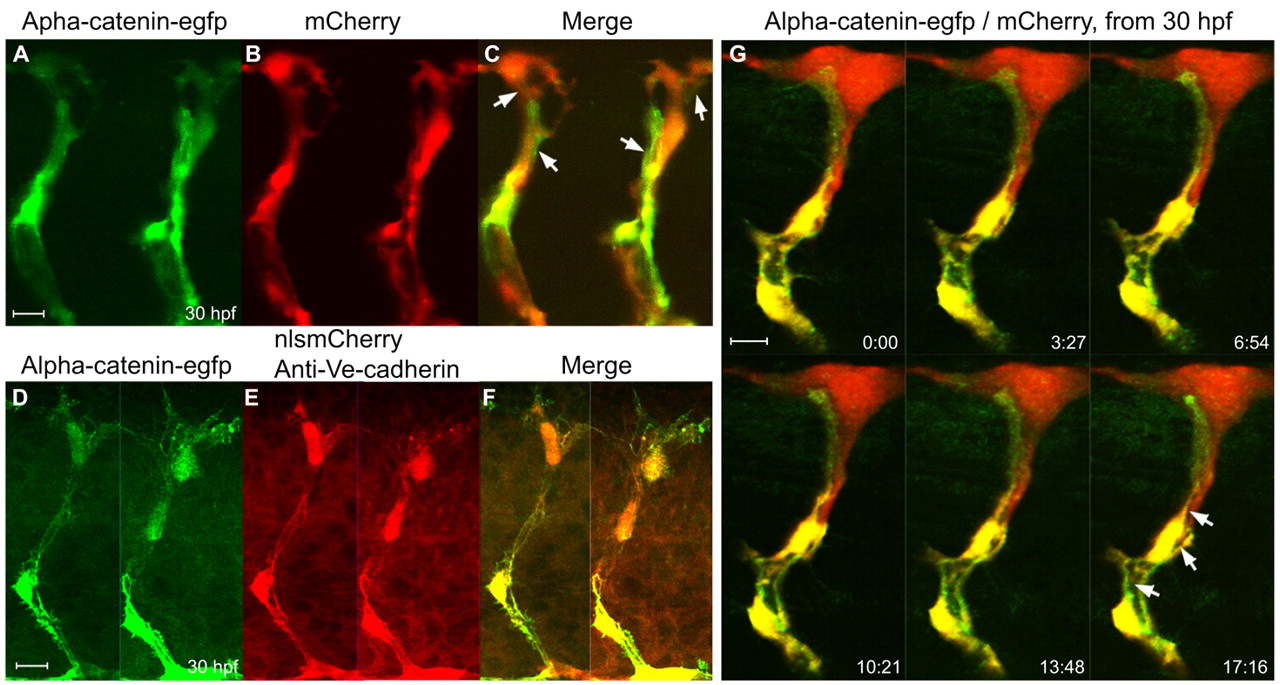
Fig. 2 α-Catenin-EGFP localizes to adherens junctions associated with the primary lumen in the ISVs. (A-C) Confocal images of the ISVs in a living Tg(flk1:α-catenin-egfp)/Tg(flk1:mCherry) zebrafish embryo at 30 hpf. Arrows in C point to long putative adherens junctions between cells. (D-F) Labeling of adherens junctions in the ISVs with anti-Ve-cadherin antibody in a Tg(flk1:α-catenin-egfp)/Tg(flk1:nlsmCherry) double-transgenic embryo. (G) Time-lapse confocal images of an ISV in a Tg(flk1:α-catenin-egfp)/Tg(flk1:mCherry) embryo showing the putative primary lumen (arrows) forming between the cellular junctions. The time format is minutes:seconds.