
|
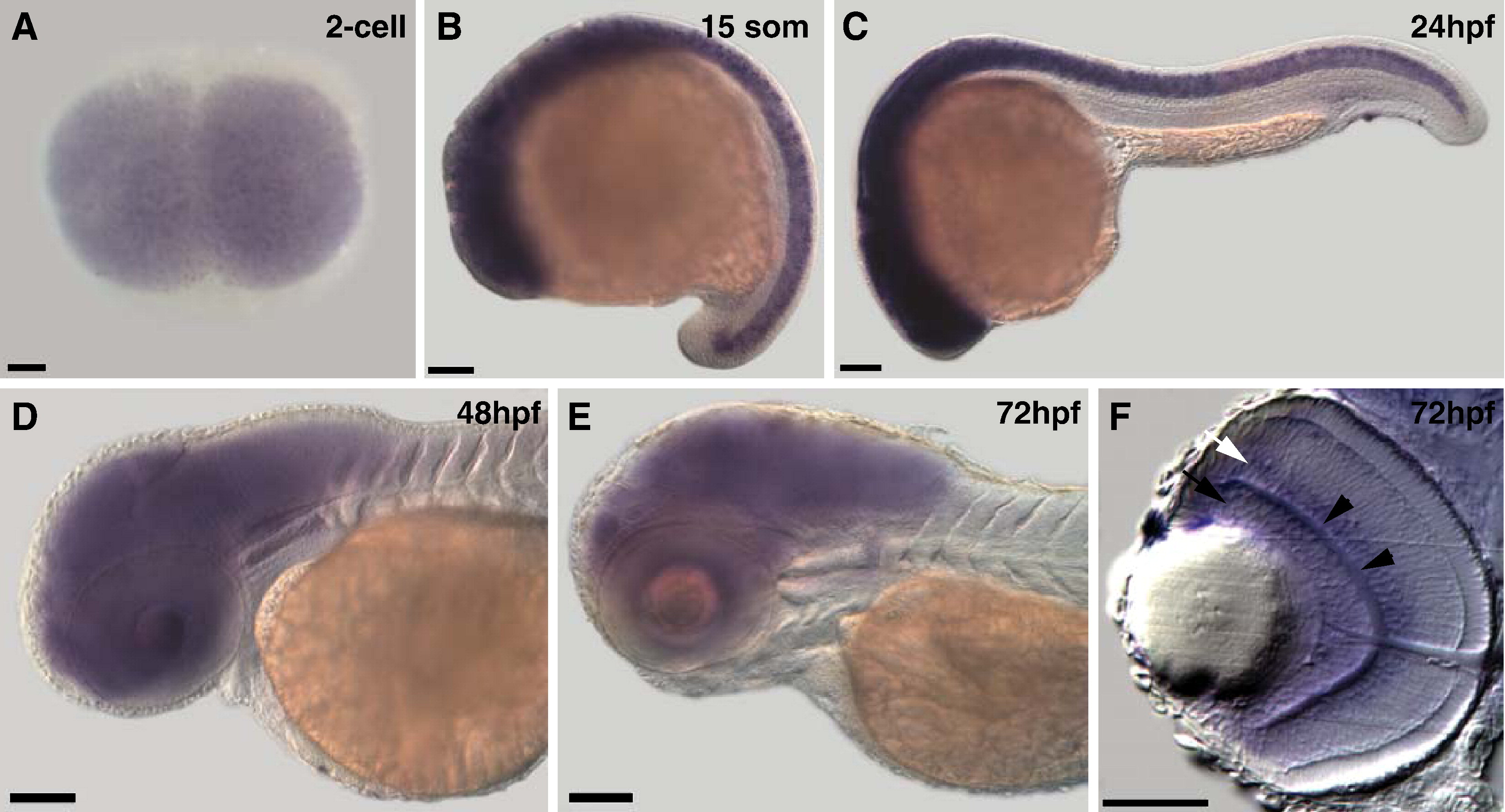
Fig. 4 cyfip2 is broadly expressed in the CNS. (A) Animal pole view of a two-cell embryo showing maternal expression of cyfip2. (B and C) Lateral views of a 15 somite and 24hpf embryo, respectively. cyfip2 is broadly expressed in the CNS. (D and E) From 48hpf to 72hpf, when axons first arrive at the optic tectum and begin to innervate it, cyfip2 is still broadly expressed in the brain and retina. (F) Coronal section through a 72hpf eye. cyfip2 is expressed in the RGC layer (black arrow), the inner plexiform layer (black arrowheads), and the inner nuclear layer (white arrow). Scale bars = 100 μm (A–E), 50 μm (F).
Reprinted from Developmental Biology, 344(2), Pittman, A.J., Gaynes, J.A., and Chien, C.B., nev (cyfip2) Is required for retinal lamination and axon guidance in the zebrafish retinotectal system, 784-794, Copyright (2010) with permission from Elsevier. Full text @ Dev. Biol.