
Fig. 6
|
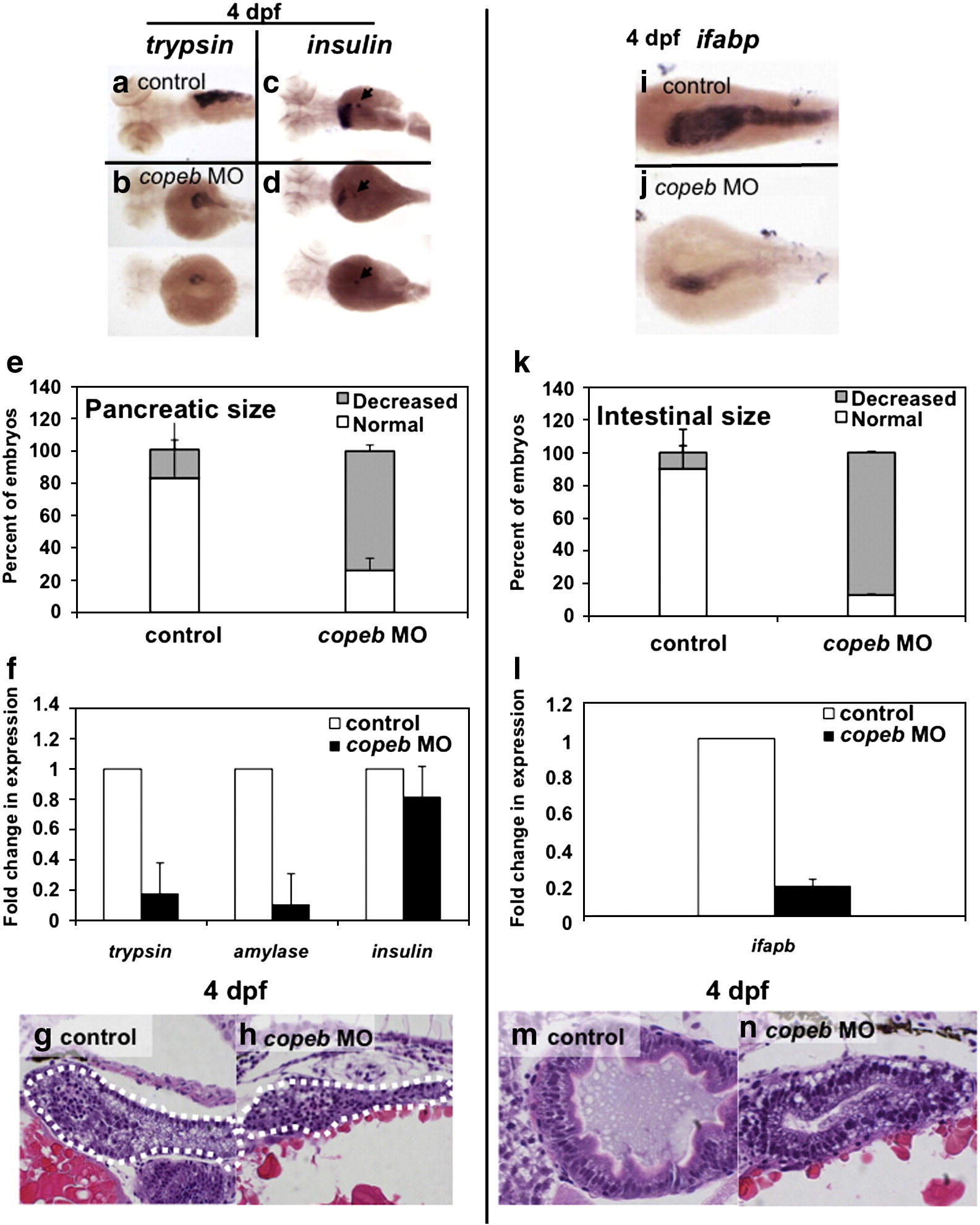
Fig. 6 copeb is required for expansion of the exocrine pancreas and intestine. Pancreatic development was assessed by WISH for trypsin (a, b) and insulin (arrowhead in c, d) in 4 dpf uninjected controls (a, c) and copeb ATG morphants (b, d). The fabp10 probe was included with the insulin probe as a control. Two morphants are shown to indicate the range of phenotype. (e) The size of the exocrine pancreas was scored as either normal, indicating a wide pancreatic head and long tail, or reduced, indicating that the trypsin stained tissue was less than ½ what is typically observed in control fish; 73.9% of the copeb morphants had reduced pancreas size versus 17.7% in controls (n = 50, p < 0.01). (f) Expression of markers of the exocrine pancreas (trypsin and amylase) and endocrine pancreas (insulin) were analyzed by qPCR, with rppO as a reference in 4 dpf morphant and control larvae. The average fold change 3 clutches of morphants were determined by normalizing to the average expression in uninjected controls. (g, h) H&E stained sagittal sections of 4 dpf embryos through the widest part of the pancreas (outlined) illustrate a well formed exocrine pancreas in control embryos and a significantly smaller exocrine pancreas in copeb morphants. (i, j) WISH using ifabp as a marker of gut epithelial cells on 4 dpf embryos revealed slight staining of only the intestinal bulb in copeb morphants. (k) 4 dpf uninjected controls and copeb morphants processed by WISH for ifabp. Controls and morphants from 3 clutches (n = 50 for each) were scored as normal if they had staining in a thick and curved intestinal bulb as well as staining in the distal intestine, or as decreased if the staining was confined to the intestinal bulb and if the intestine was thin. Decreased gut ifabp staining was observed in 10.0% of the control versus 87.1% of the morphants (p < 0.01). (l) ifabp mRNA level in 4 dpf embryos using qPCR with rppO as a reference was averaged in 3 clutches of copeb morphants and normalized to the average expression in controls. (m, n) H&E staining of 4 dpf embryos shows well-differentiated intestinal epithelial cells copeb morphants, however the intestinal villi do not form.
Reprinted from Developmental Biology, 344(1), Zhao, X., Monson, C., Gao, C., Gouon-Evans, V., Matsumoto, N., Sadler, K.C., and Friedman, S.L., Klf6/copeb is required for hepatic outgrowth in zebrafish and for hepatocyte specification in mouse ES cells, 79-93, Copyright (2010) with permission from Elsevier. Full text @ Dev. Biol.